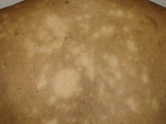
Definition
Post-inflammatory hypopigmentation refers to a reduction in skin pigmentation that occurs as a result of damage, either external or internal, following inflammation or injury. This condition causes the affected skin to appear lighter than the surrounding areas due to a disruption in melanin production, the pigment responsible for skin color.
The human skin plays a vital role in protecting the body from physical, chemical, and biological hazards. Comprising three main layers—epidermis, dermis, and hypodermis—skin also serves an aesthetic function, enhancing one's appearance. However, inflammation caused by various factors, such as infection or injury, can damage these layers, often leaving marks in the form of discoloration.
While typically benign and devoid of physical symptoms, this condition frequently presents significant cosmetic concerns, particularly for individuals with darker skin, where the contrast between the patches and natural skin tone is more pronounced. Early treatment is essential to improve this condition and prevent irreversible changes.
Causes
Post-inflammatory hypopigmentation can arise from both internal and external causes. Inflammation or injury to the skin may result in certain areas becoming visibly lighter. This discoloration occurs due to damage to the pigment-producing cells responsible for melanin production during the inflammatory process. The resulting light patches are particularly prominent in individuals with darker skin tones.
Specifically, the causes of post-inflammatory hypopigmentation include:
Internal Factors:
- Inflammatory skin conditions:
- Seborrheic dermatitis, characterized by white scales and redness on the skin and scalp
- Atopic dermatitis, inflammation caused by allergic reactions
- Contact dermatitis, inflammation resulting from exposure to specific substances
- Psoriasis, marked by thick white scales on the skin
- Infections like syphilis affecting the skin
External Factors:
- Post-treatment effects:
- Laser therapy
- Cryotherapy
- Chemical peels
- Skin patches following surgery
- Post-injury effects:
- Burns caused by chemicals
- Burns caused by heat
Risk factor
Post-inflammatory hypopigmentation can occur at any age and affects both sexes equally. However, it is more pronounced in individuals with darker skin tones due to the contrast between the lighter patches and their natural skin color. Those who have suffered burns or injuries are at greater risk of developing these patches. Surgical procedures on the skin can also contribute to the emergence of post-inflammatory hypopigmentation.
Certain skin diseases, along with specific medications, may cause pale patches in people with darker complexions. These include topical treatments containing strong steroids or acne medications that feature benzoyl peroxide.
Symptoms
Post-inflammatory hypopigmentation typically does not cause discomfort such as itching or pain in the affected areas. The only visible sign is the appearance of pale patches in regions previously affected by inflammation or injury. These patches feel smooth to the touch, with no scaling, and are noticeably lighter or more faded in color compared to the surrounding skin.
Diagnosis
Your doctor will inquire about the pale patches you are experiencing, as well as ask about factors that may have contributed to their appearance, such as any existing conditions, medications you are using, or previous infections, injuries, or surgeries. Diagnosis of post-inflammatory hypopigmentation is confirmed through a physical examination. During the examination, your doctor will observe the presence of pale or lighter patches compared to the surrounding skin. These patches, upon touch, will feel flat and non-scaly.
Supplementary Tests
In some cases, supplementary tests are required to distinguish post-inflammatory hypopigmentation from other skin conditions with similar signs. One such test is a biopsy, where a small sample of skin tissue is taken from the pale patch for microscopic examination. Additionally, a Wood's lamp examination may be employed. This test involves using a Wood's lamp to examine the lesions or pale patches under ultraviolet light, helping differentiate post-inflammatory hypopigmentation from other skin conditions that present similar symptoms.
Management
Complications
Although it typically does not present with symptoms, this condition can have psychological ramifications and affect the mental well-being of those afflicted. This is particularly true if the post-inflammatory hypopigmentation appears on highly visible areas of the body, such as the face, neck, hands, or feet. These skin patches can significantly impact the individual's appearance, potentially diminishing their quality of life.
In cases of post-inflammatory hypopigmentation, the skin contains a diminished amount of melanin, the pigment responsible for providing color. Melanin plays a crucial role in protecting the skin from the harmful effects of ultraviolet radiation from the sun. If left unaddressed, this condition may pose a risk for the development of skin cancer in the future.
Prevention
Maintain the health and protection of your skin. In cases of post-inflammatory hypopigmentation, your skin contains a reduced amount of melanin, the pigment responsible for coloration. This renders you more susceptible to skin cancer in the future, necessitating the routine application of sunscreen every day to prevent such complications. Utilize sunscreen with an SPF of 30 or higher to safeguard your skin from harmful UV radiation from the sun.
When to see a doctor?
Promptly consult your doctor if you notice multiple patches of skin that are pale or lighter than the surrounding area without any known cause. If these lighter patches appear without a prior history of injury, or if you experience numbness or a loss of sensation in the affected area, seek medical attention without delay.
Looking for more information about other diseases? Click here!
- dr. Yuliana Inosensia
Madu, P. N., Syder, N., & Elbuluk, N. (2020). Postinflammatory Hypopigmentation: A Comprehensive Review of Treatments. Journal of Dermatological Treatment, 1–13. doi:10.1080/09546634.2020.1793892
Dermcoll.edu.au. ACD A-Z of Skin - Post-inflammatory hypopigmentation. Accessed March 23, 2022, from https://www.dermcoll.edu.au/atoz/post-inflammatory-hypopigmentation/
Medlineplus.gov. (2022, January 14). Skin Pigmentation Disorders | Hyperpigmentation | MedlinePlus. Accessed March 23, 2022, from https://medlineplus.gov/skinpigmentationdisorders.html
Yenny, S. W. (2019). Common Hypopigmentation Disorders (pp. 01–20). Universitas Andalas.
Healthline.com. (2018, September 29). Hypopigmentation: Causes, Risk Factors, Treatments, More. Accessed March 23, 2022, from https://www.healthline.com/health/skin-disorders/hypopigmentation


