Definition
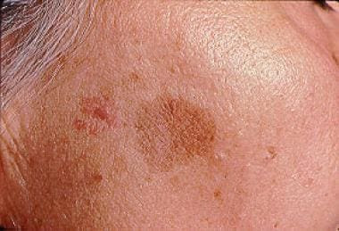
Lentigo is a pigmented patch or macule on the skin darker than the surrounding skin. It can be flat or slightly raised and has a well-defined border. Unlike freckles, lentigines do not usually fade in the winter. The term lentigo is derived from its shape, which resembles a small lentil or bean.
Lentigo can be found worldwide, with varying numbers based on the type of lentigo.
Causes
The causes of lentigo formation depend on its type, namely:
- The cause of lentigo simplex is unknown. Some cases of lentigo simplex in children have been reported following the tacrolimus cream used to treat atopic dermatitis (allergic dermatitis).
- Solar lentigo and ink spots are associated with sun exposure in fair-skinned people. It can happen because of sunburn, indoor tanning, and phototherapy, especially photochemotherapy (PUVA). Repeated exposure to ultraviolet light can trigger DNA damage leading to increased melanin production (skin pigment).
- Radiation lentigo is caused by high doses of localized radiation, which can be found, for example, in radiation therapy.
- Genetic factors may play a role in the formation of other types of lentigo, such as XP, LEOPARD syndrome, Peutz-Jeghers syndrome, and inherited patterned lentigines.
Risk factor
Lentigo can affect men and women in all age groups and races. Factors that increase the risk of developing lentigo vary depending on the types of lentigo.
- Solar lentigo caused by exposure to ultraviolet light is especially prevalent in fair-skinned people compared to dark-skinned people, as dark-skinned people have more natural skin pigments that protect them against ultraviolet exposure
- Inherited patterned lentigines can be found in black people, especially American Indians and in people who have relatives with red hair
- PUVA lentigo, tanning-bed lentigo, ink spot lentigo are more common in fair-skinned people
- PUVA lentigo is more common in men than women
- Tanning-bed lentigo is more common in women than men
Lentigo can occur in children and adults, but children are more likely to develop lentigo associated with certain syndromes. There are lentigo associated with several syndromes that appear at birth or during childhood.
Symptoms
Lentigo lesions can be singular, but more often appear as multiple lesions. Most lentigos are smaller than 5 mm in diameter. Lentigo is surrounded by normal skin. Lentigo may appear slowly over several years, or it may appear instantly. The color of lentigo can be the same or vary from brownish to black.
Lentigo is divided into different types depending on its lesions, location, contributing factors, and whether it is associated with another disease or condition. Some types of lentigo are:
- Lentigo simplex. This occurs during childhood and early adulthood. Lesions could be found on the torso and limbs. The lesions are small, brown, round, or oval in shape and appear as flat or raised lesions. The lesion borders may be regular or irregular, and the surface could be dry. Lentigo simplex may disappear with time.
- Solar lentigo. This lentigo is a precursor to seborrheic keratosis (wart-like lesions). The most commonly affected parts of the body are those frequently exposed to sunlight, such as the hands, face, and lower limbs. In addition, sunburn on the shoulders can also cause solar lentigo. The lesion's surface is dry, and the lesion edges often resemble the shape of a moth-eaten outline. Solar lentigo may enlarge slowly until the lesions become several centimeters in diameter. However, it can also disappear, often due to lichenoid keratosis. Lentigo lesions with uncommon appearances can be difficult to distinguish from early-stage melanoma (skin cancer).
- Ink spot lentigo or reticulated lentigo. There are fewer of these types of lentigos than solar lentigos. This lentigo occurs after sunburn exposure in fair-skinned people. The shape of the lesions is irregular and dark brown to black in color.
- PUVA lentigo. This lentigo is similar to ink spot lentigo but could be found after photochemotherapy (PUVA). It is found anywhere in the body areas that are exposed to PUVA.
- Tanning bed lentigo. This lentigo is similar to ink spot lentigo but occurs after indoor tanning. The location where the lesions are found is on any part of the body exposed to tanning beds.
- Radiation lentigo. These lesions appear in areas of the body that are exposed to radiation. This condition is associated with the presence of advanced-stage radiation dermatitis (radiation-induced skin inflammation).
- Melanotic macules or mucosal melanosis. These lesions appear on the surface of mucous membranes or nearby hairy skin such as the lips, vulva, penis and anus. The color of this lentigo is dark brown.
- Generalized lentigines or multiple lentigines. This type of lentigo is found on areas of the body either covered or exposed to the outside world from early childhood. Small skin lesions may merge to form larger patches. This lentigo is not associated with any particular syndrome.
- Patterned lentigines. A person with this type of lentigo has an inherited tendency to get lentigo on the face, lips, buttocks, palms and soles. This condition is especially prevalent in people of African ethnicity.
- Lentiginosis syndrome. This condition includes LEOPARD/Noonan, Peutz-Jeghers, Lugier-Hunziker, and other diseases. These syndromes are inherited and characterized by widespread lentigo on the body that are present from birth or appear in early childhood. It is associated with neural, endocrine, and mesenchymal tumors.
Diagnosis
Doctors usually diagnose lentigo by inspecting the lesion appearances on the skin. If melanoma (skin cancer) is suspected, dermatoscopy could be performed:
- Dermatoscopy is a procedure to look deeper into the skin structure
- Biopsy is the removal of tissue sample to be examined under a microscope
Management
Most cases of lentigo do not require treatment. Attempts to lighten the color of lentigo are not always successful. Here are the treatments that can be given:
- SPF 50+ broad spectrum sunscreen
- Hydroquinone bleaching cream
- Alpha hydroxy acid
- Vitamin C
- Retinoid
- Azelaic acid
- Cysteamine cream
- Peeling. Treatment of solar lentigo using chemical peels can be better than cryotherapy (cold therapy to help destroy tumors), as there are fewer side effects (side effects include pain and light patches of skin or hypopigmentation).
After a few months of use, creams that contain the above ingredients can help in lightening lentigo.
Single lesion of lentigo can be permanently removed by these treatments:
- Cryotherapy. This is a simple treatment for individual lentigo lesions. Many experts choose cryotherapy as the first choice treatment for solar lentigo. This procedure has a high success rate because melanocytes (cells that produce melanin) are sensitive to freezing with liquid nitrogen. Melanocytes will freeze at temperatures of -4 to -7°C.
- Trichloroacetic acid (TCA) 33%.
- Pigment-specific or short-pulsed lasers to destroy skin pigments in solar lentigo. This therapy can give significant improvement without any worrisome side effects.
- Intense pulsed-light (IPL) is a treatment that utilizes light waves.
Cryotherapy is more effective than TCA 33% in the treatment of solar lentigo lesions found on the back and hands, especially in fair-skinned individuals. Both therapies have the risk of post infllamatory hyperpigmentation on the treated skin.
Complications
Lentigo is a benign condition that usually persists, the lesions do not disappear easily and could increase in number with age and sun exposure. Lentigo lesions that appear on clothed skin areas protected from the sun may fade and disappear in time.
Especially in women, lentigo lesions can be non-aesthetically pleasing.
Prevention
Lesions of lentigo associated with ultraviolet radiation can be prevented by protecting yourself from sun exposure. Covering the body with clothes prevents new lentigo lesions more effectively than sunscreen. In addition, women should also be careful in using tanning beds.
When to see a doctor?
You should consult a doctor if you notice any changes in the lentigo lesions, such as changes in size, shape, or color, and if the lesions start to feel itchy or bleed.
Looking for more information about other diseases? Click here!
- dr. Yuliana Inosensia
Lentigo: Causes, Features, and Treatment — DermNet. Dermnetnz.org. (2022). Retrieved 3 May 2022, from https://dermnetnz.org/topics/lentigo.
Lentigo Treatment & Management: Medical Care, Surgical Care, Prevention. Emedicine.medscape.com. (2022). Retrieved 3 May 2022, from https://emedicine.medscape.com/article/1068503-treatment#d9.
Gangguan Kosmetik Karena Kelainan Pigmentasi Kulit (Cosmetic Disorders Due to Skin Pigmentation Abnormalities). Jurnal.ugm.ac.id. (2022). Retrieved 3 May 2022, from https://jurnal.ugm.ac.id/bik/article/view/4645.


