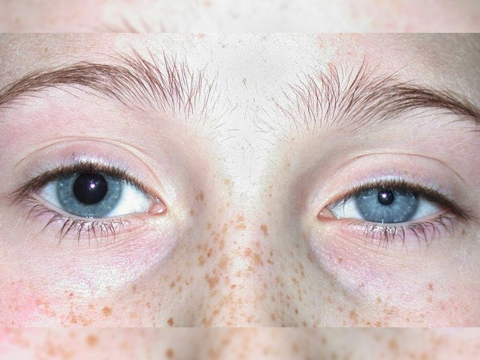
Definition
The pupil is the dark part of your eye surrounded by the iris, the part that gives your eye its color. The pupil functions as a pathway for light to enter the eye, allowing humans to see objects in front of them. The size of the pupil changes based on the brightness of the room you are in. In a dark room, your pupil will dilate to allow more light into the eye, enabling you to see in the dark. Conversely, in a bright room, your pupil will constrict to limit the amount of light entering the eye, preventing glare. Both pupils respond to changes in light simultaneously—if your right pupil constricts, your left pupil will constrict as well, and vice versa. Therefore, the condition of your pupils and how they respond to light can provide clues to your doctor about your current health status.
Anisocoria
Anisocoria is a condition where one pupil's diameter is larger than the other. The term anisocoria comes from Greek: "aniso-" meaning unequal, "kore" meaning pupil, and "ia" meaning abnormal condition. About 1 in 5 healthy individuals have this condition. A difference in pupil diameter of about 0.5–1 mm is considered normal. One of the most famous examples is the singer David Bowie, whose left pupil was larger than his right due to an accident. However, if the pupil diameter is more than 1 mm or remains permanently unequal, it may be an early sign of a more serious disease.
Causes
Anisocoria can occur due to the following mechanisms:
- There is an obstruction preventing one pupil from constricting, making the other pupil appear larger.
- An obstruction prevents one pupil from dilating. These issues are caused by pupils' ability to dilate and constrict. Another cause of anisocoria is a disorder in the muscles that regulate pupil diameter. Any factor that disrupts the balance of regulation or directly affects the muscles can cause anisocoria.
Risk factor
The causes of anisocoria range from physiological or normal variations to more serious conditions. These include:
- Normal variation (20% of the population)
- Use of asthma medications
- Post-cataract surgery
- Brain aneurysm
- Brain hemorrhage following head trauma
- Brain tumor or abscess
- Increased intraocular pressure in one eye
- Glaucoma
- Increased intracranial pressure due to brain swelling, brain hemorrhage, or stroke
- Meningitis or brain infection
- Migraine
- Seizure
- Diabetic neuropathy
- Horner syndrome due to carotid artery dissection
- Nerve paralysis
Symptoms
Anisocoria is characterized by a significant difference in the size of the pupils. A size difference of 3-5 mm between the eyes is considered significant. The most common symptom is vision disturbance. Other symptoms depend on the underlying cause of anisocoria.
Diagnosis
To diagnose anisocoria, your doctor will determine if your condition is truly anisocoria and differentiate between normal and abnormal anisocoria by examining your pupils in both dark and bright environments and observing their response to light changes. If necessary, your doctor may administer 1–2 drops of 4–10% cocaine to both eyes and examine them 30–45 minutes later. Apraclonidine 0.5–1% and pilocarpine 0.1% can also be used to detect anisocoria. Other possible examinations include lab tests related to syphilis and imaging of the head, neck, and chest to detect Horner syndrome or cranial nerve VII paralysis.
Management
Treatment for anisocoria depends on the underlying condition:
- Physiological or normal anisocoria does not require further treatment.
- Anisocoria caused by trauma may require surgery to repair structural damage.
- Anisocoria due to eye conditions such as glaucoma or infection, can be treated with medication.
- Anisocoria resulting from drug use can be managed by reducing exposure to those drugs.
- Anisocoria related to nerve paralysis may require collaboration between an ophthalmologist and a neurologist.
- Anisocoria caused by life-threatening conditions such as stroke, hemorrhage, or vascular abnormalities needs prompt treatment.
Complications
Anisocoria rarely causes significant complications, though some may occur. A pupil with a larger diameter can lead to visual aberrations due to excessive light entering the eye (glare). A pupil with a smaller diameter can make it difficult to see due to limited light entry. Complications from anisocoria arise from the underlying disease causing the condition.
Prevention
Anisocoria itself cannot be prevented, so prevention focuses on the underlying diseases that may cause it.
When to see a doctor?
If you experience changes in pupil diameter after head trauma, seek immediate medical attention. If you notice a newly developed difference in pupil size, it may indicate a serious condition. Immediate medical attention is required if you experience additional symptoms, such as:
- Blurred or complete loss of vision
- Double vision
- Increased sensitivity to light
- Fever
- Headache
- Nausea and vomiting
- Eye pain
- Stiff neck
It is important to note that anisocoria is a clinical sign, not a primary disease. A comprehensive examination is necessary to identify the main disease causing anisocoria.
Looking for more information about other diseases? Click here!
- dr. Yuliana Inosensia
Encyclopedia, M. (2021). Anisocoria: MedlinePlus Medical Encyclopedia. Retrieved 31 October 2021, from https://medlineplus.gov/ency/article/003314.htm
Payne WN, Blair K, Barrett MJ. Anisocoria. [Updated 2021 Aug 22]. In: StatPearls [Internet]. Treasure Island (FL): StatPearls Publishing; 2021 Jan-. Available from: https://www.ncbi.nlm.nih.gov/books/NBK470384/.
Anisocoria - EyeWiki. (2021). Retrieved 31 October 2021, from https://eyewiki.aao.org/Anisocoria.
Anisocoria Clinical Presentation: History, Physical, Causes. (2021). Retrieved 31 October 2021, from https://emedicine.medscape.com/article/1158571-clinical#b5.


