Definition
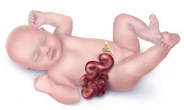
Gastroschisis is a congenital defect characterized by abnormalities in the abdominal wall. Consequently, the baby's intestines protrude from the body and pass through a cavity in the abdomen. The cavity may vary in size, ranging from small to large, and might include organs such as the stomach and liver, in addition to the intestines, which are located outside the infant's body. This disorder has a prevalence of 1 in 4,000 live births.
Causes
The failure of abdominal wall closure can be classified into two distinct conditions: omphalocele and gastroschisis. Omphalocele is characterized by a protective layer of the umbilical cord and the peritoneum, which covers the intestine protruding from the stomach. In gastroschisis, the intestines protrude from the body without a protective layer.
The etiology of gastroschisis remains incompletely understood. Gastroschisis occurs due to a defect in the formation and growth of the front body wall during the embryonic stage. Consequently, the stomach wall weakens, leading to the perforation of the wall and the protrusion of the intestine from the body. Research conducted on mice indicates that insufficient levels of folic acid, oxygen deprivation, and the use of salicylic products contribute to the poor development of the abdominal wall. Nevertheless, these findings may not necessarily correspond to those observed in humans.
Risk Factor
Several factors contribute to the development of gastroschisis, including:
- Infants born to pregnant women under 20
- Alcoholic and smoking mothers
- The prevalence of gastroschisis in male infants is 1.5 times higher than in female infants
Symptoms
The condition known as gastroschisis is easily identifiable. Part of the intestines often exit the body through a hole in the stomach, which allows food and water to enter the body. The hole is typically around 5 cm in diameter and situated to the right of the umbilical cord. The intestines can appear normal with their curvature, but swelling can also cause them to appear enormous. This swelling may result from the inability to see other intestinal anomalies, such as an obstruction in another section of the intestine.
Diagnosis
A gastroschisis diagnosis can be confirmed at approximately 20 weeks or 5 months into a pregnancy. The previously mentioned defect is detectable using ultrasonography. An ultrasound observation of perforation on the right umbilical cord and a protruding loop of the intestine outside the uterine cavity helps diagnose gastroschisis. Exposure of the intestines to amniotic fluid may increase their thickening and enlargement. Fetuses with gastroschisis may be born with a low birth weight. Furthermore, unsafe conditions such as fetal mortality or premature spontaneous birth can manifest within the uterus during pregnancy. Regular ultrasound examinations will be performed throughout pregnancy to assess the viability of the intestine.
Laboratory analyses may also be performed. Elevated levels of alpha-fetoprotein (AFP) are indicative of gastroschisis.
Following delivery, an ultrasound can be performed to identify abnormalities in the intestines outside the stomach. The ultrasonography examination can detect an obstruction in the intestines. It is also possible to tell if the abdominal wall can be stitched directly or if a "silo" technique needs to be used by looking at the inflammation, the peristaltic movement of meconium (fetal waste) through the intestines, and the properties of the intestinal fluids. The silo serves to coat the intestines during ongoing inflammation. After the intestines have shown improvement, the treatment and control of gastroschisis can be performed.
A prenatal ultrasound can also examine multiple organs, such as the heart and lungs, to identify concurrent abnormalities. Chromosome analysis can be conducted to identify genetic abnormalities responsible for gastroschisis.
Management
Gastroschisis management is categorized into four different stages:
- Maternity. Throughout pregnancy, regular ultrasound examinations are used to monitor the fetus, as it is typical for fetuses with gastroschisis to exhibit low birth weight. Additional examinations can be conducted to verify the viability of the fetus and the potential for preserving the intestines located outside the abdomen.
- Delivery. Vaginal delivery is possible for a gastroschisis baby. The results of fetal screening, ultrasound, and lung development affect gestational age, determining when to deliver the baby. For optimal outcomes, it is advisable to deliver a fetus diagnosed with gastroschisis at a referral hospital, as the management of the condition typically necessitates interdisciplinary cooperation. Given this condition, there is a risk that the baby will be delivered prematurely.
- Initial treatment. After the infant's birth, management procedures persist in the treatment room. The air evaporation rate from the external environment is approximately 2.5 times higher than the evaporation rate from other typical body areas. Therefore, the intestines that are located outside the body will be contained within a silo. Silos serve the dual purpose of safeguarding the intestines and enhancing blood circulation and oxygen supply to the intestines. A gastric tube may be placed to reduce gastric pressure. The infant will also get intravenous (IV) antibiotics to prevent infection and deliver fluids as required. The stability of the infant's condition will be protected and maintained.
- Surgical treatment. It is worth considering a surgical intervention. The primary objective of surgery is to reposition the intestines within the body while minimizing the occurrence of intestinal damage or an excessive rise in intra-abdominal pressure. Two surgical procedures might be considered for this condition: primary repair and delayed closure. Essentially, these two operations will alleviate inflammation in the intestines that come into contact with amniotic fluid and facilitate its entry into the stomach. This procedure is performed concurrently with assessing the intestine's intended insertion site. This allows for prompt identification and subsequent treatment of obstructions or perforations in the intestine. Typically, the intestines can be introduced into the stomach within a week. Nevertheless, restoring intestinal function typically takes approximately 4-6 weeks, resulting in a diminished ability for the infant to metabolize breast milk adequately during this period.
Complications
There are an extensive number of complications associated with gastroschisis. Fetuses afflicted with gastroschisis have an increased risk of preterm birth by 28%. Furthermore, due to impaired intestinal function, nutrition will be administered intravenously, significantly increasing the risk of infection. Additionally, infection of the gastroschisis closure scar and intestinal inflammation may ensue. In addition, infants afflicted with gastroschisis frequently manifest additional intestinal complications, including obstructions, perforations, and inflammation of the intestines. These complications may necessitate the implementation of an intestinal transplant for the infant.
Prevention
It is possible to prevent gastroschisis through careful pregnancy planning. Pregnancy should ideally occur when a woman is at least 20 years old, as this significantly reduces the risk of developing complications such as gastroschisis. Aside from that, pregnant women should stop smoking and excessive alcohol consumption.
When to See a Doctor?
If your infant exhibits the presence of protruding intestines, regardless of the presence or absence of a protective covering, it is essential to seek medical attention immediately. Gastroschisis significantly elevates the susceptibility to intestinal infection, perhaps leading to mortality. If gastroschisis is detected after birth, an immediate referral to a specialized hospital will typically be prepared. Prenatal detection of gastroschisis helps with birth preparation in locations with comprehensive resources.
Looking for information about other diseases? Click here!
- dr. Yuliana Inosensia
Facts about Gastroschisis | CDC. (2020). Retrieved 18, 2022, from https://www.cdc.gov/ncbddd/birthdefects/gastroschisis.html
Glasser, J. (2019). Pediatric Omphalocele and Gastroschisis (Abdominal Wall Defects): Background, Pathophysiology, and Etiology. Retrieved 18, 2022, from https://emedicine.medscape.com/article/975583-overview
Rentea, R., & Gupta, V. (2021). Gastroschisis. Retrieved January 18, 2022, from https://www.ncbi.nlm.nih.gov/books/NBK557894/


