Definition
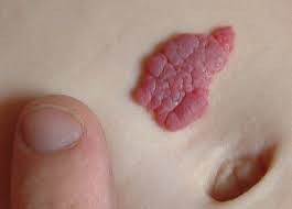
A capillary hemangioma is a benign (non-cancerous) tumor formed due to the abnormal growth of capillaries (small blood vessels). It is the most common type of hemangioma, appearing as a bright red lump.
Capillary hemangiomas are typically not present at birth but become noticeable within the first six months of life. They grow rapidly during infancy and may spontaneously resolve as the child ages. Generally, these hemangiomas grow quickly until the age of 12 months and begin to shrink between 12 months and 5 years. They usually disappear completely by the age of 5 or 6.
Capillary hemangiomas can arise in various areas of the body. When located near the eye, they may appear on the eyelids, conjunctiva, or within the eye’s orbit.
Causes
Capillary hemangiomas result from an overgrowth of the cells lining blood vessels. These cells divide abnormally, forming new tissue that appears as a tumor, which is benign in nature. However, the precise cause or trigger for this abnormal growth remains unknown.
In capillary hemangiomas of the eye, there are two phases of tumor growth. The first phase, the rapid growth (proliferative) phase, usually occurs between 8 and 18 months of age, marked by an increase in the cells stimulating blood vessel growth. After this proliferative phase ends, the next phase of gradual tumor shrinkage (involution) begins. During this phase, the increased cells from the first phase return to normal levels. Approximately 50% of tumors shrink by the age of 5, and 75% by the age of 7.
Risk factor
Genetic or hereditary factors have not been shown to significantly increase the risk of capillary hemangiomas in children. Some studies have reported that capillary hemangiomas are rare in African races. Premature birth and low birth weight have been associated with a higher risk of developing capillary hemangiomas after birth.
Other suspected risk factors include female gender, with a ratio of hemangioma cases in girls to boys ranging from 3:2 to 5:1. Developmental disturbances during pregnancy may also increase the risk of capillary hemangiomas in children.
Symptoms
Capillary hemangiomas typically appear after birth, within the first six months of life (30% are present at birth, 50% occur in children aged 1-2 months, and 90% by six months). These benign tumors can appear on the skin, beneath the skin, or around the eyes.
Symptoms vary from patient to patient. Generally, capillary hemangiomas present as raised, bright red patches or lumps, firm to the touch and with well-defined edges. These red lumps can arise on various parts of the body. They blanch when pressed, and their texture is similar to a sponge.
In the eye area, hemangiomas appear as red spots growing near the eye. If they affect the eyelids, the child may gradually have difficulty opening them. Patients typically present with lumps on the eyelids or eyebrows. If the hemangioma extends into the eye socket, the eyeball may protrude, potentially impairing vision.
Amblyopia, or lazy eye, is found in 43-60% of patients with eyelid hemangiomas. Anisometropia, or unequal refractive power between the eyes, may be detected during an eye exam, resulting from corneal pressure caused by the hemangioma. Strabismus (crossed eyes) can also occur if the hemangioma compresses the eye, affecting muscle function.
Although hemangiomas may resolve on their own, they often leave a persistent discoloration in the area where the lump has subsided, though this is usually less intense than when the hemangioma first appeared.
Diagnosis
Physicians diagnose capillary hemangiomas based on symptom evaluation, patient medical history, physical examination, and supporting tests. Laboratory tests such as immunohistochemical staining may aid diagnosis, often revealing a positive result for factor VIII, a protein involved in blood clotting.
Though hemangiomas are typically visible on the skin, an ultrasound is necessary to determine whether the tumor has spread to nearby tissues or organs. Radiological exams are also valuable for diagnosis. Ultrasound imaging shows an irregularly contoured lump. If the hemangioma is near the eye and suspected to extend into the eye socket, a CT scan or MRI may be conducted.
A CT scan will show the tumor as a mass with low blood flow, without affecting surrounding tissue or bone erosion. The lump becomes more visible when a contrast agent is injected into the blood vessels. On MRI, capillary hemangiomas appear as solid tumors with well-defined borders. Microscopic examination of tissue samples reveals an overgrowth of the cells lining blood vessels.
Management
Capillary hemangiomas can be treated in various ways depending on their location, severity, and whether they cause vision problems. Hemangiomas typically take years to shrink. The affected skin may remain reddish, slightly wrinkled, or appear normal, depending on the stage of healing.
Medications
Propranolol, a beta-blocker used for heart conditions, is now the first-line treatment for hemangiomas. It is administered orally and is safe for use. Heart rate and blood pressure monitoring are sometimes necessary at the start of treatment, which may require a short hospital stay. In some cases, beta-blocker eye drops can help if the hemangioma near the eye is small.
Steroids may also be prescribed to halt hemangioma growth. Depending on the tumor’s size and location, steroids can be taken as tablets, injected directly into the hemangioma, or applied topically. Steroids may have unwanted side effects.
Other Procedures
Laser therapy may be used to treat surface hemangiomas, helping prevent growth, reduce size, or lighten the color. Traditional surgery to remove hemangiomas near the eye is typically performed for small, well-defined tumors located beneath the skin.
Complications
While most capillary hemangiomas shrink over time, some grow rapidly and may compress surrounding tissues or organs. Hemangiomas near the eye can cause:
- Amblyopia (lazy eye)
- Proptosis (protrusion of the eyeball)
- Keratitis
- Compression of the optic nerve
- Eye ulcers and bleeding
Steroid treatment must be carefully monitored to prevent side effects, such as:
- Low blood pressure
- Slowed heart rate
- Low blood sugar
- Airway narrowing
- Sleep disturbances
- Diarrhea
- Electrolyte imbalances
Surgical complications may include damage to surrounding tissues.
Prevention
Since the exact cause of hemangiomas is unknown, there is no effective way to prevent capillary hemangiomas. Fortunately, they usually resolve on their own. Consult your doctor about suitable treatments to prevent worsening symptoms and complications.
When to see a doctor?
If your baby exhibits symptoms of a capillary hemangioma, as described above, seek medical attention for proper evaluation and treatment. Early diagnosis and intervention can prevent complications such as compression of surrounding organs or tissues.
Looking for more information about other diseases? Click here!
- dr. Yuliana Inosensia
https://emedicine.medscape.com/article/1218805-overview
Capillary hemangioma. (2020). Retrieved 22 April 2022, from https://aapos.org/glossary/capillary-hemangioma
Koka K, Patel BC. (2021). Capillary infantile hemangiomas. Retrieved 22 April 2022, from https://www.ncbi.nlm.nih.gov/books/NBK538249/
Osigian, CJ. (2021). Capillary hemangioma. Retrieved 22 April 2022, from https://eyewiki.aao.org/Capillary_Hemangioma#Demographics


