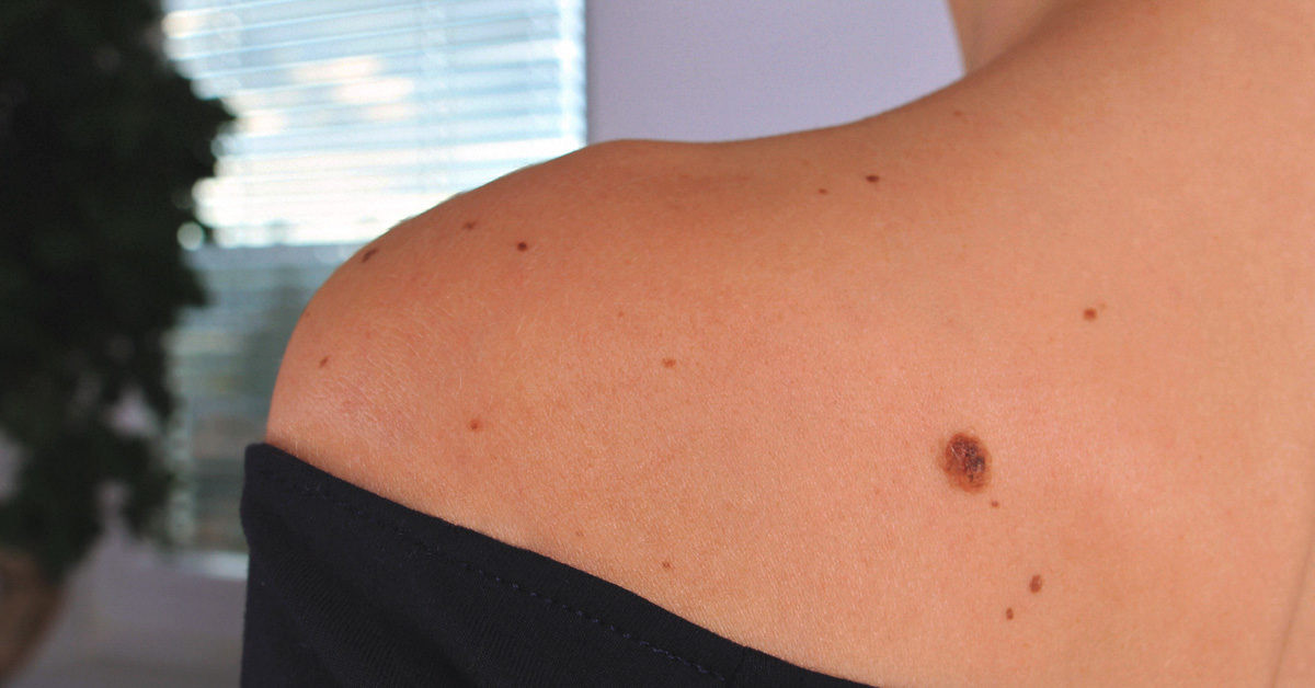
Definition
A nevus, also known as a mole or a melanocytic nevus, is a common type of skin lesion. Moles are typically small, appearing as dark brown spots or areas, and are caused by a group of cells called melanocytes, which produce skin pigment. Generally, during childhood and adolescence, as many as 10-40 moles can be present on the skin's surface, and these can change with age.
Most moles are harmless and rarely become cancerous. However, you need to regularly check your moles because they could develop into skin cancer cells, especially malignant melanoma.
Almost everyone has at least one melanocytic nevus on the surface of their skin. Approximately 1% of individuals are born with one or more melanocytic nevi at birth (congenital moles). Individuals with fair skin tend to have more melanocytic nevi than individuals with darker skin. Melanocytic nevus acquired as a child or adult can be affected by sun exposure and can disappear on its own.
Causes
The cause of mole appearance is currently not known with certainty. There are no significant genetic or environmental factors that contribute to the growth of the nevus. However, there are several studies which state that there are several factors such as location in the body, environmental factors and factors of nevus evolution that play a role in the emergence of a mole in a person. There is also a suspicion that dysplastic nevus is inherited genetically and passed down through the family (in an autosomal dominant manner).
The growth of moles is caused by cells in the skin, also known as melanocytes, growing in a group together. Generally melanocyte cells are scattered along the skin surface. These cells produce skin color called melanin (a natural pigment that gives color to the surface of an individual's skin).
Risk factor
Risk factors for the appearance of moles are sometimes influenced by genetic factors or environmental factors. Melanocytic nevi are generally benign so they can disappear on their own in certain cases. Pale skin and age can be other risk factors that could trigger the development of melanocytic nevus.
What you need to be careful of is if you have risk factors where an existing nevus or mole becomes melanoma (skin cancer). Some risk factors for melanoma include having a job or working in places with high exposure to ultraviolet rays from sunlight, having a family history of melanoma, low economic status (poverty), and low knowledge about melanoma.
Melanoma is a skin cancer usually characterized by moles that change shape to become irregular, increase in size by more than 6 millimeters, the edges of the moles are not clear, the mole has various colors and sometimes bleeds easily.
Symptoms
Symptoms and signs of moles usually appear as small brown spots or bumps. However, moles can vary in color, shape or size. Moles can be brown, black, blue, red or pink with a flat or protruding texture and are sometimes accompanied by hair growth. Most moles are oval and less than 6 millimeters in size.
Moles can appear from birth which are known as congenital nevus and can be larger than usual, they could also be found on the face or limbs.
Diagnosis
The diagnosis of a nevus, or mole, can be established through medical interview, physical examination by finding a mole or a distinctive brown to black spot, and diagnostic test with dermoscopy to help and further ensure that the existing mole is a benign growth.
You should inform your doctor if you or your family member has previously had melanoma (skin cancer) because moles can become cancerous if you have several risk factors for melanoma. Information such as when symptoms first appeared will also help doctors diagnose and determine the best treatment when the patient is first examined by a doctor.
If the doctor suspects that a mole is malignant or has the potential to become cancerous, the mole will be surgically removed and sent to the laboratory to be examined under a microscope.
Management
Until now there are no particular medicines to treat moles. In general, moles are benign lumps or tumors. If you feel it to be unsightly, the doctor will perform surgery to remove the mole using several therapies that need to be consulted and discussed together. If an atypical nevus is found, the doctor will perform a biopsy first and then carry out surgery.
Complications
Nevus generally does not have any special complications. However, minor surgical procedures during a biopsy or small specimen or tissue removal by surgery can trigger complications, such as infection or bleeding.
Another complication that can arise is melanoma. Some people who have moles have a higher risk of developing melanoma (skin cancer). Some risk factors that can increase the risk of melanoma are being born with a large nevus with the size of 5 cm, having more than 50 nevus, having a large and irregularly shaped nevus known as atypical or dysplastic nevus.
Prevention
Prevention is needed to avoid complications that can arise from a nevus, namely melanoma. Several steps can be taken to detect early changes of a nevus to melanoma. Here are the steps you can take:
- Pay attention to any changes in your moles by doing a self-examination of your skin every month, the things you need to assess are their shape, boundaries, color or size.
- Protect your skin from ultraviolet rays exposure with sunscreen containing at least SPF 30
- If you don't have sunscreen, you can use physical objects that block sunlight such as umbrellas or
Apart from the steps above, if you have never seen a doctor, you should do a general examination and if you find a condition or disease that can increase your risk of developing melanoma, the doctor will recommend starting treatment as early as possible before the condition becomes more severe and worse.
When to see a doctor?
If you find that your mole has undergone significant changes such as increasing in size over time, bleeding easily, the shape of the mole becomes irregular, the color of the mole changing or is not the same as other moles on the other side of your body, you should consult a dermatologist. The doctor will conduct a medical interview, physical examination, and certain diagnostic tests if needed to determine a definite diagnosis and appropriate treatment.
Looking for more information about other diseases? Click here!
- dr. Yuliana Inosensia
Bodman MA, Al Aboud AM. Melanocytic Nevi. [Updated 2022 May 1]. In: StatPearls [Internet]. Treasure Island (FL): StatPearls Publishing; 2022 Jan-. Available from: https://www.ncbi.nlm.nih.gov/books/NBK470451/
Medscape. Melanocytic nevi. November 2019. https://emedicine.medscape.com/article/1058445-overview#a4
Dermnet NZ. Melanocytic naevus. January 2016. https://dermnetnz.org/topics/melanocytic-naevus
Mayo Clinic. Moles. May 2022. https://www.mayoclinic.org/diseases-conditions/moles/symptoms-causes/syc-20375200


