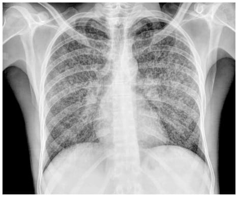
Definition
Pleural effusion refers to the abnormal accumulation of fluid on the outer side of the lungs, contained within the pleura—a thin membrane lining the lungs and chest wall. The pleura's two layers create a space normally filled with a small amount of fluid, facilitating lung movement within the chest during breathing. This fluid can move into and out of the pleura through the bloodstream and lymphatic system.
Causes
Pleura effusion can be caused by various conditions. These conditions can cause problems both in the pleura, blood vessels, and lymphatic channels. Common causes include:
- Leakage from other organs. Leakage from other organs is often seen in congestive heart failure, liver diseases, and kidney diseases, resulting in fluid accumulation that may seep into the pleural space.
- Cancer. Cancer, particularly lung cancer, as well as cancers that metastasize to the lungs or pleura.
- Infection. Infections that often cause pleura effusion are tuberculosis infections (which are caused by the bacteria Mycobacterium tuberculosis) and pneumonia (pulmonary conditions due to infection with viruses, bacteria, fungi, or parasites).
- Autoimmune conditions. Autoimmune conditions like systemic lupus erythematosus (SLE) and rheumatoid arthritis, where the body's immune system mistakenly attacks its own cells, contributing to pleural effusion.
- Problems with pulmonary blood vessels. Pulmonary blood vessel problems, including pulmonary embolism, where a blood vessel in the lungs becomes blocked suddenly.
Pleural effusion is categorized into transudate and exudate:
- Transudate: characterized by fluid accumulation with a composition similar to normal pleural fluid. Usually caused by fluid leakage through intact pleura, common in congestive heart failure.
- Exudate: Formed from blood, protein, inflammatory cells, and bacteria leaking from damaged blood vessels. Often requires drainage depending on fluid volume and inflammation severity. Common causes include pneumonia and lung cancer.
Risk Factor
The risk of pleural effusion increases based on underlying conditions predisposing to its development. For instance, individuals with a history of heart attacks or strokes are at elevated risk of pleural effusion due to congestive heart failure. In women, pleural effusion often arises from breast cancer, reproductive organ malignancies, and lupus. Conversely, in men, asbestos exposure, chronic pancreatitis (stemming from prolonged alcohol consumption), and rheumatoid arthritis are common risk factors.
Symptoms
Pleural effusion may remain asymptomatic with small fluid volumes but typically manifests symptoms with moderate to large effusions or concurrent inflammation. Common symptoms include:
- Shortness of breath or wheezing
- Chest pain, particularly during deep breaths (pleuritic chest pain)
- Fever
- Cough
Symptoms may vary depending on the underlying cause of the pleural effusion.
Diagnosis
Diagnostic evaluation of pleural effusion involves investigating potential causes. A thorough medical history, including hepatitis or alcohol abuse, aids in identifying liver or pancreas-related effusions. Physical examination may reveal clues to underlying conditions like heart failure, malignancies, or liver or kidney disorders.
Imaging studies, such as X-rays or Computed Tomography (CT) scans, may be conducted based on facility availability. Thoracentesis, involving fluid extraction from the pleura with ultrasound guidance, may be performed if fluid accumulation is significant.
The laboratory will test this fluid to determine if it is a transudate or an exudate. If the fluid is an exudate, it will be utilized for cell counting, Gram staining, and culture (to identify the bacteria infecting the lungs or pleura). Other laboratory tests can be used to investigate the autoimmune etiology of pleural effusion. In addition, pleural tissue can be obtained if tuberculosis or cancer is suspected.
Management
The treatment of pleural effusion is dependent on the underlying cause. If pleural effusion persists and causes severe difficulty breathing, the fluid can be evacuated by thoracentesis. This thoracentesis, however, has a maximum fluid volume restriction. If too much fluid is extracted during thoracentesis, the lungs may swell or produce a hole in the chest cavity (pneumothorax).
If pleural effusion occurs as a result of malignancy (lung, breast, etc.), the doctor may recommend pleurodesis. Pleurodesis is a technique that aims to attach the pleura on the chest wall to the pleura on the lungs, resulting in no interpleural space. Pleurodesis is used to keep fluid from accumulating in the interpleural area, allowing the patient to feel more comfortable.
Aside from that, another option is to insert a chest tube to continuously drain fluid from the body. To avoid clogging, clean this hose every 6–8 hours. Diet can also help with certain types of pleural effusions, such as those caused by fat accumulation. In this case, you can restrict your intake of fatty meals to reduce pleural effusion.
Treatment for pleural effusion might also include medication. These drugs will be administered according to the cause of the pleural effusion. For example, if a pleural effusion arises due to heart failure, medication will be administered to allow fluid to flow back into the blood vessels. Meanwhile, if pleural effusion is caused by an infection, medications will be administered to treat the infection.
Complications
Complications of pleural effusion vary depending on the underlying cause. Effusions stemming from relatively benign conditions like lupus or rheumatoid arthritis may resolve spontaneously. Conversely, effusions secondary to cancer typically carry a poorer prognosis, with survival rates ranging from 12 to 24 months. Untreated effusions can lead to respiratory distress and pleural infections.
Prevention
Preventing pleural effusion entails addressing the underlying causes of effusion. For instance, mitigating the risk of heart failure involves managing heart disease risk factors through regular exercise, reducing fat intake, increasing fiber consumption, and adhering to prescribed medications for existing conditions like hypertension or diabetes. Similarly, preventing liver and pancreatic diseases entails abstaining from alcohol and avoiding illicit drug use involving syringes.
When to See a Doctor?
Seek medical attention if you experience worsening shortness of breath during exertion or while lying down, significant weight loss, or persistent coughing. These symptoms may signify underlying conditions contributing to pleural effusion, such as heart failure, cancer, or tuberculosis. Early consultation facilitates prompt identification and management of the underlying cause, potentially reducing the risk of recurrence.
- dr. Alvidiani Agustina Damanik
Boka, K. (2021). Pleural Effusion: Background, Anatomy, Etiology. Retrieved 18 February 2022, from https://emedicine.medscape.com/article/299959-overview
Hoffman, M. (2020). Pleural Effusion. Retrieved 18 February 2022, from https://www.webmd.com/lung/pleural-effusion-symptoms-causes-treatments
Krishna, R., & Rudrappa, M. (2021). Pleural Effusion. Retrieved 18 February 2022, from https://www.ncbi.nlm.nih.gov/books/NBK448189/


