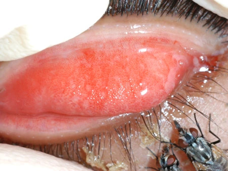
Definition
Dacryocystitis refers to the inflammation of the tear sac, which typically occurs due to a blockage of the tear duct leading to the nose, resulting in the accumulation of tears in the sac. The condition is most common in two age groups: newborns and adults over 40 years of age. Among newborns, congenital tear duct obstruction affects approximately 6%, with dacryocystitis occurring in 1 out of every 3,884 live births. In adults, this condition is more frequently observed in women than in men.
Causes
Tears are produced by the lacrimal glands located in the upper outer region of the eyelid, functioning to moisturize and soothe the eyes. These tears are then directed through channels into the tear sac, and subsequently drained into the nasal cavity.
In most cases, dacryocystitis results from an obstruction in the duct responsible for transporting tears into the nasal cavity. Dacryocystitis can be classified based on its duration (acute or chronic) and its onset (congenital or acquired). Acute dacryocystitis is often caused by bacterial infections, including Staphylococcus aureus, Streptococcus beta hemolyticus, Staphylococcus epidermidis, Streptococcus pneumoniae, and Pseudomonas aeruginosa. Chronic dacryocystitis, on the other hand, is caused by long-standing blockages, recurrent infections, or autoimmune disorders such as systemic lupus erythematosus (SLE).
Congenital dacryocystitis is generally attributed to incomplete development of the tear ducts, leading to an inability to drain amniotic fluid from the tear ducts, which then become infected by bacteria. In adulthood, dacryocystitis may develop due to age-related narrowing of the tear ducts, trauma such as nasal fractures, nasal surgery, tumors, or treatments for other conditions like glaucoma.
Risk Factor
The risk factors for dacryocystitis are closely linked to its causes. In terms of gender, women are more prone to the condition due to their narrower tear ducts compared to men. Age is also a significant risk factor, as the risk increases with age due to the narrowing of the tear duct, which slows down the flow of tears. Nasal conditions such as a deviated septum (a bending of the nasal cavity partition) and nasal inflammation can also elevate the risk of dacryocystitis. Trauma or surgical procedures involving the nasal bone further increase susceptibility. Additionally, tumors, autoimmune diseases, and dacryoliths—accumulations of cells, fat, and debris within the tear duct—may also contribute to duct blockages, heightening the risk. Glaucoma treatments, particularly with medications such as timolol and pilocarpine, are also associated with an increased risk of dacryocystitis.
Symptoms
The symptoms of dacryocystitis vary depending on whether the condition is acute or chronic. In acute dacryocystitis, symptoms may develop over a few hours to days and typically include pain, redness, and swelling in the region between the eyes and nose. The pain is usually confined to this area, and redness may extend to the bridge of the nose. Additionally, a pus-like discharge may be expelled from the end of the duct along with tears. In contrast, chronic dacryocystitis is often characterized by excessive tearing and pus production. Fever may also be present, along with visual complaints, such as reduced visual acuity. However, decreased vision is generally due to an abnormal tear layer caused by the blockage.
Diagnosis
The diagnosis of dacryocystitis is typically established by a physician through inquiry about the disease's progression and patient history. In cases of acute dacryocystitis, the doctor may apply gentle pressure to the tear duct to extract some fluid for bacterial testing, such as Gram stain and culture. If there is a history of trauma or surgery, the doctor might suggest a CT scan. Additionally, imaging techniques like dacryocystography (DCG) can be utilized if a structural abnormality in the tear duct is suspected. If conditions like rhinitis or narrowing of the nasal cavity are present, nasal endoscopy may be performed to determine whether these are contributing factors to dacryocystitis.
An evaluation using dye may also be conducted to assess tear flow. A duct obstruction is suspected if the dye remains in place for more than five minutes.
In chronic dacryocystitis, the doctor may assess for systemic diseases like SLE or Wegener's granulomatosis (inflammation of the blood vessels in the nose, throat, lungs, and kidneys). Antibody tests such as Antinuclear Antibody (ANA) for SLE and Antineutrophilic Cytoplasmic Antibody (ANCA) for Wegener’s granulomatosis can also be performed.
Management
Treatment for dacryocystitis varies depending on the cause. In cases of acute dacryocystitis, a physician may massage the tear ducts gently to promote tear drainage. The application of warm compresses to the swollen area may also be recommended to alleviate inflammation. Oral antibiotics may be prescribed to address bacterial infections. For congenital dacryocystitis, parents may be instructed to gently massage the area from the center of the eye towards the nose to facilitate tear drainage. If signs of inflammation such as redness or swelling appear, antibiotics may also be prescribed. This condition often resolves within six months to a year with these methods.
Chronic dacryocystitis, on the other hand, typically requires surgical intervention. The surgery is aimed at reopening the tear ducts to restore normal tear drainage into the nasal cavity.
Complications
Complications from dacryocystitis can arise due to prolonged inflammation or infection. Persistent inflammation may lead to the formation of abnormal tear ducts, known as fistulas. Infections can result in the formation of pus-filled cavities (abscesses) or may spread to the brain's membranes, potentially causing meningitis. Other severe complications include blockage of the blood vessels in the cavernous sinus region of the head, blindness, and even death.
Prevention
Preventing dacryocystitis involves maintaining proper hygiene in the nasal cavity and eyelids. Nasal cleanliness can be achieved through the use of nasal sprays to encourage the flow of tears into the nose. Eyelid hygiene can be maintained with warm compresses and regular cleaning.
When to see a doctor?
It is advisable to see a doctor if sudden swelling occurs in the area between the eyes and nose, whether or not it follows trauma or surgery. If a newborn exhibits swelling in this area, medical attention should be sought for further examination and appropriate therapy.
Looking for more information about other diseases? Check here, yes!
- dr. Yuliana Inosensia
Alsuhaibani, A., Goldstein, T., Vargason, C., Goel, S., Barmettler, A., & Yen, M. (2021). Dacryocystitis - EyeWiki. Retrieved 4 November 2021, from https://eyewiki.aao.org/Dacryocystitis.
Gilliland, G. (2019). Dacryocystitis: Practice Essentials, Background, Epidemiology. Retrieved 4 November 2021, from https://emedicine.medscape.com/article/1210688-overview#a4.
Taylor, R., & Ashurst, J. (2021). Dacryocystitis. Retrieved 4 November 2021, from https://www.ncbi.nlm.nih.gov/books/NBK470565/.


