Definisi
Koledokolitiasis atau yang dikenal juga dengan batu saluran empedu adalah terbentuknya satu atau lebih batu empedu di saluran empedu. Batu saluran empedu dapat berasal dari kantung empedu atau terbentuk langsung di saluran empedu.
Saluran empedu atau common bile duct adalah saluran kecil dari hati yang membawa cairan empedu dari kantung empedu ke usus halus. Cairan empedu ini berperan dalam pencernaan lemak. Kantung empedu adalah organ berbentuk seperti buah pir yang terletak di bawah hati atau liver, di sisi kanan atas perut. Batu-batu ini biasanya bisa menetap di dalam kantung empedu atau bisa keluar melalui saluran empedu. Batu tersebut biasanya terbuat dari pigmen empedu, kalsium, atau kolesterol.
Adanya batu pada saluran empedu dapat menyebabkan cairan empedu tidak bisa mengalir ke usus halus dan dapat kembali ke organ hati. Menurut penelitian, sekitar 1-15% orang dengan batu empedu juga akan mengalami batu saluran empedu atau koledokolitiasis.
Penyebab
Berdasarkan bahan pembentuk batu, ada dua jenis batu empedu, yaitu batu empedu pigmen dan batu empedu kolesterol. Batu empedu kolesterol merupakan jenis batu empedu yang paling umum dan sering terlihat dengan warna kuning. Batu kolesterol disebabkan oleh empedu yang mengandung terlalu banyak kolesterol, terlalu banyak bilirubin (pigmen kuning hasil penguraian sel darah merah), dan tidak cukupnya garam empedu.
Batu kolesterol dapat terjadi jika kantung empedu tidak cukup sering atau tidak sepenuhnya mengosongkan kantungnya. Batu kolesterol biasanya ditemukan pada pasien obesitas yang jarang olahraga atau pasien yang sengaja menurunkan berat badan dengan cepat.
Batu pigmen ada yang berupa batu pigmen hitam, sebagian besar tersusun dari pigmen saja, dan ada yang berupa batu pigmen coklat yang tersusun dari campuran pigmen dengan lemak empedu. Penyebab terbentuknya batu pigmen masih belum diketahui. Batu pigmen biasanya terbentuk pada orang yang memiliki:
- Sirosis hati (terdapatnya jaringan parut pada hati)
- Infeksi saluran empedu, bakteri menjadi sumber pigmen coklat pada batu saluran empedu
- Menerima pemberian nutrisi melalui pembuluh darah atau infus saja (pada pasien yang dirawat di rumah sakit dalam waktu lama)
- Pasca tindakan reseksi usus atau pengangkatan sebagian usus
- Kelainan darah yang diturunkan, menyebabkan hati membuat terlalu banyak bilirubin
Batu saluran empedu juga dapat dipicu oleh kolangitis, yaitu peradangan kantung empedu. Kantung empedu yang sudah membaik dari peradangan akan membentuk jaringan parut. Jaringan parut menyebabkan cairan empedu menggenang dan mengendap di dalam kantung empedu.
Berdasarkan letak asal dan mekanisme terbentuknya, batu saluran empedu dibedakan menjadi:
- Batu primer, biasanya berupa batu pigmen coklat yang terbentuk di saluran empedu
- Batu sekunder, biasanya berupa batu kolesterol, yang terbentuk di kantung empedu dan berpindah ke saluran empedu
- Batu residu, yaitu batu yang tertinggal saat prosedur pengangkatan kantung empedu yang dilakukan kurang dari 3 tahun terakhir
- Batu berulang, terjadi pada saluran yang sudah dioperasi lebih dari 3 tahun
Faktor Risiko
Orang-orang dengan riwayat batu empedu atau penyakit kantung empedu berisiko terkena batu saluran empedu. Bahkan mereka yang kantung empedunya telah diangkat juga dapat mengalami kondisi koledokolitiasis, terjadi pada sekitar 4,6-18,8% kasus. Ada faktor risiko yang bisa diperbaiki, dan tidak bisa diubah. Selama faktor risiko yang bisa diperbaiki ini dikurangi dengan merubah gaya hidup, risiko terkenanya penyakit ini bisa berkurang. Hal-hal berikut ini dapat meningkatkan risiko terkena batu saluran empedu, yaitu:
- Obesitas
- Diet rendah serat, tinggi kalori, tinggi lemak
- Kehamilan
- Puasa berkepanjangan
- Penurunan berat badan yang cepat
- Kurang aktivitas fisik
- Kadar kolesterol darah yang tinggi
Faktor risiko yang tidak dapat diubah meliputi:
- Usia, kelompok dewasa tua biasanya memiliki risiko lebih tinggi terkena batu empedu
- Jenis kelamin wanita lebih mungkin memiliki batu empedu
- Etnis, yaitu orang Asia, Indian-Amerika, dan Meksiko-Amerika memiliki risiko lebih tinggi memiliki batu empedu
- Riwayat keluarga, faktor genetik diperkirakan memiliki peran terjadinya batu saluran empedu
Gejala
Batu di saluran empedu bisa tidak bergejala selama berbulan-bulan atau bahkan bertahun-tahun. Namun, jika batu tersangkut dan menghalangi aliran saluran empedu, dapat muncul beberapa gejala berikut:
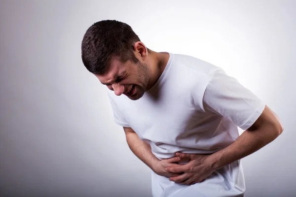
- Nyeri perut hebat terutama pada daerah kanan atas atau tengah atas perut
- Demam
- Kuning pada kulit atau area putih (sklera) mata
- Nafsu makan menurun
- Mual dan muntah
- Tinja berwarna seperti tanah liat
Rasa nyeri yang disebabkan oleh batu saluran empedu bisa hilang-timbul atau bertahan lama. Rasa nyeri awalnya dapat bersifat ringan yang kemudian memberat. Nyeri yang berat mungkin memerlukan penanganan medis darurat. Terkadang, pasien menyalah-artikan rasa nyeri berat tersebut sebagai serangan jantung.
Diagnosis
Jika mengalami gejala batu saluran empedu, dokter akan memastikan apakah benar terdapat batu di saluran empedu. Dokter dapat menggunakan salah satu dari pemeriksaan pencitraan berikut:
- USG perut, merupakan prosedur pencitraan yang menggunakan gelombang suara berfrekuensi tinggi untuk memeriksa hati, kantung empedu, limpa, ginjal, dan pankreas
- CT scan perut
- USG endoskopi perut (EUS), pada pemeriksaan ini probe USG disisipkan pada selang endoskopi yang lentur dan dimasukkan melalui mulut untuk memeriksa saluran pencernaan
- Endoscopic retrograde cholangiography (ERCP) dapat mengidentifikasi batu, tumor, dan penyempitan saluran empedu
- Magnetic resonance cholangiopancreatography (MRCP) atau MRI kantung empedu, saluran empedu, dan saluran pankreas
- Percutaneous transhepatic cholangiogram (PTCA), merupakan pencitraan sinar-X untuk saluran empedu
Dokter juga dapat melakukan satu atau lebih pemeriksaan darah berikut untuk memeriksa kemungkinan terjadinya infeksi, lalu bagaimana kondisi fungsi hati dan pankreas. Pemeriksaan tersebut antara lain:
- Darah tepi lengkap
- Bilirubin
- Enzim pankreas
- Tes fungsi hati seperti SGOT dan SGPT
Tata Laksana
Fokus terapi adalah untuk menghilangkan penyumbatan yang terjadi di saluran empedu. Penanganan yang dapat dilakukan antara lain:
- Ekstraksi (mengeluarkan) batu
- Pemecahan batu (litotripsi)
- Pengangkatan kantung empedu dan batu sekaligus (kolesistektomi)
- Operasi penyayatan saluran empedu untuk mengeluarkan batu atau membantu batu melewati saluran empedu (sphincterotomy)
- Pemasangan stent atau ring pada saluran empedu
Terapi yang paling umum dilakukan adalah biliary endoscopic sphincterotomy (BES). Jika batu tidak keluar dengan sendirinya atau tidak dapat diangkat dengan BES, prosedur litotripsi dapat menjadi pilihan terapi selanjutnya. Prosedur ini akan memecah batu sehingga pecahannya yang berukuran lebih kecil dapat melewati saluran dengan mudah.
Pasien batu saluran empedu dan batu empedu juga dapat diterapi dengan mengangkat kantung empedu. Saat operasi, dokter akan memeriksa saluran empedu untuk memeriksa batu empedu yang tersisa.
Jika batu tidak dapat dikeluarkan sepenuhnya atau Anda tidak ingin kantung empedu Anda diangkat, dokter akan memasang stent untuk membuka saluran empedu. Tindakan ini dapat melancarkan saluran empedu dan membantu mencegah kekambuhan. Selain itu, stent juga dapat mencegah infeksi.
Komplikasi
Ketika batu tersangkut di saluran empedu, hal ini bisa menyebabkan infeksi empedu. Bakteri dari infeksi dapat menyebar dengan cepat dan berpindah ke hati. Keadaan ini dapat menyebabkan infeksi yang mengancam jiwa. Kemungkinan komplikasi lainnya adalah sirosis bilier, kolangitis atau peradangan pada saluran empedu, dan pankreatitis yang juga merupakan peradangan namun terjadi pada pankreas. Sirosis bilier adalah pembentukan jaringan parut pada hati akibat aliran empedu yang tidak lancar. Pada keadaan infeksi berat, bisa terjadi sepsis atau penyebaran infeksi ke seluruh tubuh melalui aliran darah.
Pencegahan
Jika Anda memiliki batu saluran empedu, ada kemungkinan Anda akan mengalami kekambuhan. Bahkan jika kantung empedu sudah diangkat, risiko kekambuhan akan tetap ada. Perubahan gaya hidup seperti rutin berolahraga intensitas sedang, diet tinggi serat, serta mengurangi konsumsi lemak jenuh dapat mengurangi kemungkinan Anda terkena batu saluran empedu di kemudian hari.
Kapan Harus ke Dokter?
Konsultasi ke dokter jika:
- Mengalami nyeri perut, dengan atau tanpa demam, dan tidak diketahui penyebabnya
- Mengalami jaundice atau kuning pada mata atau kulit
- Memiliki gejala koledokolitiasis lainnya seperti yang telah disebutkan di atas
Mau tahu informasi seputar penyakit lainnya? Cek di sini, ya!
- dr Hanifa Rahma
Choledocholithiasis. (2017). Retrieved 10 January 2022, from https://www.healthline.com/health/choledocholithiasis
McNicoll CF, Pastorino A, and Farooq U. (2020). Choledocholithiasis. StatPearls LC Publishing. Retrieved 10 January 2022, from https://www.ncbi.nlm.nih.gov/books/NBK441961/
Baiu I, Hawn MT. (2018). Choledocholithiasis. JAMA. 2018;320(14):1506. doi:10.1001/jama.2018.11812
Choledocholithiasis. (2021). Retrieved 10 January 2022, from https://medlineplus.gov/ency/article/000274.htm
Lindenmeyer C. (2021). Choledocholithiasis and cholangitis. Retrieved 14 January 2022, from https://www.msdmanuals.com/professional/hepatic-and-biliary-disorders/gallbladder-and-bile-duct-disorders/choledocholithiasis-and-cholangitis


