Definisi
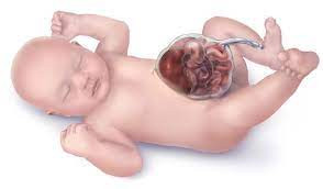
Omfalokel atau eksomfalos adalah suatu kelainan bawaan lahir pada dinding perut bayi dimana organ seperti usus, hati, atau organ dalam perut lainnya keluar dari rongga perut melalui celah pada pusar. Organ-organ ini dibungkus oleh kantung yang tipis dan transparan yang disebut sebagai lapisan peritoneum. Kantung ini bisa terbuka atau robek. Pada omfalokel, bisa keluar sebagian kecil usus saja atau dapat juga sebagian besar usus hingga banyak organ yang keluar dari rongga perut.
Sekitar 1 dari 4.200 bayi lahir dengan omfalokel di Amerika Serikat. Kebanyakan bayi dengan omfalokel memiliki cacat lahir yang lain seperti cacat pada jantung, tabung saraf, tulang, saluran cerna, saluran kemih, bibir sumbing, dan kelainan kromosom akibat mutasi genetik atau sindrom yang dapat memengaruhi anggota badan, membuat anak mengalami kesulitan belajar, gangguan mental atau perilaku. Sindrom-sindrom tersebut antara lain sindrom Beckwith-Wiedemann, sindrom Prader Willi, sindrom Patau, sindrom Edwards, dan sindrom Down
Penyebab
Pada minggu ke enam sampai sepuluh kehamilan, organ dalam perut janin mengalami pertumbuhan yang pesat sehingga ukurannya akan semakin panjang dan besar, sampai akhirnya rongga perut tidak mampu menampungnya. Organ tersebut (hati, kandung kemih, lambung, usus, limpa, indung telur, testis) lalu menonjol masuk ke tali pusat. Pada minggu ke sebelas kehamilan, rongga perut sudah cukup besar untuk usus dan hati sehingga organ tersebut normalnya masuk kembali ke rongga perut. Jika organ-organ tersebut gagal berputar dan tidak masuk kembali ke dalam perut, maka kondisi ini disebut dengan omfalokel.
Umumnya penyebab omfalokel belum diketahui dengan pasti. Beberapa bayi menderita omfalokel karena adanya kelainan pada gen atau kromosomnya. Omfalokel juga dapat disebabkan oleh kombinasi dari faktor genetik dan faktor lain seperti keadaan ibu saat kehamilannya. Ketika hamil, apa yang dikonsumsi ibu termasuk makanan, minuman, dan obat-obatan akan memengaruhi janin.
Faktor Risiko
Walaupun penyebab pastinya masih belum diketahui, diduga ada beberapa faktor yang dapat meningkatkan kemungkinan suatu bayi menderita omfalokel, yaitu:
- Usia ibu yang kurang ideal dan rentan terjadi kehamilan berisiko (di bawah 20 tahun atau di atas 40 tahun)
- Bayi dari ibu yang rutin mengonsumsi alkohol dan perokok berat lebih mungkin menderita omfalokel
- Bayi dari ibu yang mengonsumsi obat-obatan tertentu selama kehamilan, contohnya adalah obat antidepresan golongan SSRI (selective serotonin-reuptake inhibitors) seperti sertralin, sitalopram, esitalopram, atau fluoksetin
- Bayi dari ibu yang kelebihan berat badan atau obesitas baik sebelum maupun saat hamil
- Riwayat keluarga dengan omfalokel
- Ras kulit hitam
- Bayi laki-laki
- Kehamilan kembar
Gejala
Janin dengan omfalokel dapat terdeteksi dengan pemeriksaan pencitraan USG (ultrasonografi), sedangkan pada bayi yang sudah lahir, adanya omfalokel dapat dilihat secara langsung pada garis tengah rongga perut. Ukuran dari omfalokel tergantung dari seberapa banyak organ yang keluar, yaitu:
- Ukuran kecil jika hanya beberapa bagian dari usus yang keluar
- Berukuran besar jika mengandung beberapa organ perut
- Sangat besar jika celah di perut berukuran 5 cm atau lebih yang disertai dengan keluarnya organ hati
Diagnosis
Omfalokel dapat didiagnosis mulai dari kehamilan dan setelah bayi lahir. Saat hamil terdapat beberapa pemeriksaan skrining atau pemeriksaan prenatal untuk mengidentifikasi adanya kelainan bawaan lahir dan gangguan lainnya. Janin dengan omfalokel dapat memperlihatkan hasil yang tidak normal saat dilakukan pemeriksaan, antara lain:
- Pada pemeriksaan darah, terdapat peningkatan kadar alpha-fetoprotein (AFP) pada darah ibu
- Pemeriksaan ultrasonografi (USG) pada akhir trimester satu juga dapat menunjukkan adanya omfalokel
Jika saat kehamilan sudah terdeteksi adanya omfalokel, maka perlu dilakukan pemeriksaan lebih lanjut seperti:
- Pemeriksaan ultrasonografi jantung bayi
- Pengambilan cairan ketuban dari rahim dilakukan untuk mencari adanya kelainan genetik yang biasanya menyertai omfalokel, dan pemeriksaan lainnya yang ditentukan dokter
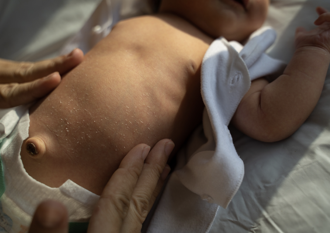
Setelah lahir, adanya omfalokel dapat langsung dilihat dengan kasat mata dengan adanya organ yang keluar dari rongga abdomen yang tertutup oleh kantung atau membran. Membran inilah yang membedakan dengan penyakit lain yaitu gastroshcisis, dimana keluarnya organ terjadi akibat tidak sempurnanya pembentukan dinding perut. Pada gastroschisis, organ tidak tertutup membran dan lokasi keluarnya organ biasanya lebih ke kiri dari garis tengah rongga perut.
Tatalaksana
Biasanya, bila saat bayi lahir ditemukan adanya omfalokel, hal pertama yang dapat dilakukan pada bayi dengan omfalokel adalah menutup organ yang keluar dengan kasa steril atau minimal kain bersih untuk menjaga kehangatan bayi dan mencegah penguapan cairan. Hindari menutup organ dengan kain yang terlalu tebal. Pilihan terapi untuk bayi dengan omfalokel adalah dengan operasi, yang dilakukan berdasarkan pertimbangan beberapa faktor yang ada di bawah ini.
- Ukuran omfalokel
Jika ukurannya relatif kecil, atau hanya sebagian kecil usus yang berada di luar rongga perut, biasanya omfalokel dapat dioperasi dalam 72 jam setelah bayi lahir. Tujuan dari prosedur operasi adalah untuk mengembalikan usus tersebut ke dalam rongga perut dan menutup celah tempat keluarnya usus dari perut.
Namun, jika ukuran omfaloker sangat besar atau banyak organ yang berada di luar perut, operasi biasanya dilakukan secara bertahap dalam waktu beberapa hari sampai beberapa minggu. Organ yang masih berada di luar akan ditutup oleh penutup khusus dan akan dimasukan satu-persatu secara perlahan. Celah keluarnya organ akan ditutup setelah semua organ masuk ke dalam perut.
Alasan prosedur operasi dilakukan secara bertahap adalah untuk keselamatan bayi. Rongga perut bayi dengan omfalokel sangat kecil dan tidak berkembang degnan sempurna untuk dapat menampung seluruh organ dalam satu waktu. Jika dipaksakan, organ yang dipaksa masuk tidak akan mendapatkan aliran darah yang cukup.
- Adanya kelainan bawaan lahir atau abnormalitas kromosom lain
- Usia kehamilan ibu saat bayi dilahirkan
Komplikasi
Komplikasi omfalokel dapat terjadi sejak proses melahirkan, dimana lapisan peritoneum yang menutupi organ pecah. Jika omfalokel berukuran sangat besar dan organ hati berada di luar, maka saat proses melahirkan organ hati dapat mengalami kerusakan.
Setelah melewati proses melahirkan komplikasi masih dapat terjadi. Oleh karena organ yang seharusnya berada dalam perut menjadi berada di luar, bayi dengan omfalokel akan memiliki banyak masalah, seperti:
- Rongga perut yang seharusnya menjadi tempat organ tersebut berada bisa tidak tumbuh ke ukuran yang normal akibat tidak ada organ di dalamnya
- Infeksi, yang menjadi masalah terutama jika kantung yang melingkupi organ tersebut terbuka atau robek
- Terkadang organ tersebut dapat tertekan atau terpeluntir (terutama usus) yang mengakibatkan aliran darah menuju organ tersebut kurang lancar. Jika aliran darah kurang lancar maka lama kelamaan akan menyebabkan kerusakan pada organ.
- Terhambatnya perkembangan paru
Suatu penelitian menyebutkan bahwa pada bayi dengan omfalokel yang berukuran besar, 23% bayi mengalami kematian dengan infeksi berat sebagai penyebab terbanyak kematian. Infeksi terjadi terutama bila kantung omfalokel terbuka.
Pencegahan
Terdapat beberapa cara yang dapat dilakukan untuk mencegah omfalokel. Secara umum adalah dengan menjaga kesehatan ibu terutama saat hamil. Ibu yang sedang hamil harus melakukan pemeriksaan kehamilan secara rutin di fasilitas kesehatan. Hindari konsumsi alkohol dan kebiasaan merokok, serta untuk selalu menjaga berat badan tubuh yang ideal.
Kapan harus ke Dokter?
Bayi dengan ofmalokel harus dibawa ke fasilitas kesehatan untuk mendapat penanganan yang tepat dan pemeriksaan lebih lanjut terkait sindrom lain yang mungkin menyertai bayi.
Setelah bayi diperbolehkan pulang ke rumah, waspadai tanda-tanda dimana bayi perlu dibawa ke dokter lagi, seperti:
- Frekuensi BAB berkurang
- Tidak bisa makan
- Demam
- Muntah warna kehijauan
- Pembengkakan area perut
- Perubahan perilaku bayi
- dr Hanifa Rahma
Facts about Omphalocele | CDC, Centers for Disease Control and Prevention. (2022). Retrieved January 20, 2022, from https://www.cdc.gov/ncbddd/birthdefects/omphalocele.html.
Pediatric Omphalocele and Gastroschisis (Abdominal Wall Defects) Clinical Presentation: History and Physical Examination. Emedicine.medscape.com. (2022). Retrieved January 20, 2022, from https://emedicine.medscape.com/article/975583-clinical.
Zahouani, T., & Mendez, M. (2022). Omphalocele. Ncbi.nlm.nih.gov. Retrieved January 20, 2022, from https://www.ncbi.nlm.nih.gov/books/NBK519010/.
Encyclopedia, M. (2022). Omphalocele: MedlinePlus Medical Encyclopedia. Medlineplus.gov. Retrieved January 20, 2022, from https://medlineplus.gov/ency/article/000994.htm.
Omphalocele: Treatment, Diagnosis, Causes, & Outlook. Cleveland Clinic. (2022). Retrieved January 20, 2022, from https://my.clevelandclinic.org/health/diseases/10030-omphalocele.


