Definisi
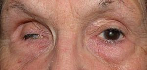
Phthisis bulbi merupakan kerusakan parah yang terjadi pada bola mata. Kerusakan ini disebut sebagai “tahap akhir” atau end-stage eye dari kerusakan mata. Phthisis bulbi dapat disebabkan oleh banyak hal, mulai dari peradangan luas pada mata hingga komplikasi dari operasi mata. Bola mata yang mengalami phthisis bulbi akan menjadi lebih kecil dibandingkan bola mata normal akibat proses penyusutan. Mata yang mengalami phthisis bulbi tidak dapat berfungsi dengan baik karena terjadi berbagai perubahan pada struktur bola mata. Kondisi ini merupakan indikasi operasi mata.
Penyebab
Terdapat beberapa kelompok penyebab dari phthisis bulbi, antara lain:
- Trauma/kecelakaan pada bola mata, seperti trauma tajam, trauma tumpul, dan trauma akibat bahan kimia basa.
- Infeksi, seperti keratitis, endoftalmitis, panoftalmitis.
- Ablasio retina, kondisi lepasnya lapisan retina dari mata.
- Pasca operasi, seperti operasi katarak, glaucoma, kornea, vitrectomy, dan lain-lain.
- Peradangan jangka panjang, seperti uveitis.
- Kelainan vascular, seperti retinopathy of prematurity (ROP)–kondisi kebutaan pada bayi prematur yang disebabkan oleh pemberian oksigen terlalu tinggi pada saat resusitasi; dan retinopati diabetes.
- Kelainan kongenital atau bawaan lahir.
- Tumor, seperti retinoblastoma.
- Obat, seperti cidofovir.
Semua penyebab tersebut dapat menyebabkan ocular hypotony atau penurunan tekanan bola mata, kerusakan pembuluh darah mata, dan peradangan terus menerus. Penurunan tekanan bola mata hingga di bawah 5 mmHg, dapat menghambat penghantaran nutrisi dan oksigen yang dibutuhkan oleh mata yang semakin mengganggu struktur bola mata. Jika kerusakan struktur sudah sangat lanjut, phthisis bulbi dapat terjadi.
Faktor Risiko
Memiliki penyakit yang berhubungan dengan peradangan jangka panjang pada tubuh dapat meningkatkan risiko untuk mengalami phthisis bulbi, terutama akibat peradangan pada struktur mata bagian dalam. Penyakit tersebut antara lain:
- Psoriasis
- Arthritis rheumatoid
- Ankylosing spondylitis (arthritis pada tulang belakang)
- Kolitis ulseratif
- Herpes
- AIDS
- Penyakit Kawasaki
- Sifilis
- Tuberkulosis
- Retinoblastoma pada Anak, beberapa laporan menunjukkan bahwa phthisis bulbi merupakan presentasi dari retinoblastoma yang sudah terlambat ditangani
Jika Anda memiliki kondisi tersebut atau mengalami penyakit autoimun, lakukan pemeriksaan mata secara berkala untuk mendeteksi perubahan bola mata lebih dini.
Gejala
Karena phthisis bulbi merupakan kondisi yang berangsur-angsur, gejala yang dirasakan akan semakin buruk seiring berjalannya waktu. Gejala yang akan dialami antara lain:
- Pandangan buram
- Floaters, melihat bayangan hitam bergerak melewati mata
- Sensitif terhadap cahaya
- Mata nyeri
- Mata merah
- Mata bengkak
- Kebutaan
- Mata menjadi lebih kecil dibandingkan mata yang sehat
Jika mengalami phthisis bulbi, mata akan mengalami penyusutan dan bagian putih dari mata atau sklera akan menebal.
Diagnosis
Dokter Anda akan bertanya mengenai riwayat penyakit dan perjalanan penyakit dari kondisi phthisis bulbi Anda. Pemeriksaan fisis akan dilakukan menggunakan oftalmoskopi atau slit lamp untuk melihat kondisi bola mata Anda secara lebih detail. Kemudian, dokter akan melakukan pemeriksaan radiologis, seperti CT scan atau MRI untuk melihat penyebab utama dari kondisi tersebut, seperti keganasan, pertumbuhan tulang, dan lain-lain. Pemeriksaan darah juga dapat dilakukan untuk menentukan penyebab. Terdapat beberapa penyakit yang dapat memiliki tampilan seperti phthisis bulbi, seperti mata yang kecil dan atrofi (penyusutan mata akibat sebab lain).
Tatalaksana
Jika phthisis bulbi belum terjadi, dokter mata Anda akan merekomendasikan untuk menatalaksana penyakit yang mendasari terlebih dahulu dengan menggunakan steroid untuk penyakit autoimun dan antibiotik untuk penyakit infeksi.
Jika phthisis bulbi telah terjadi, pengobatan bertujuan untuk meredakan nyeri pada bola mata dan perbaikan kosmetik. Pengobatan tidak bertujuan untuk mengembalikan penglihatan. Operasi merupakan satu-satunya cara untuk menangani phthisis bulbi. Mata yang mengalami phthisis bulbi dapat dikeluarkan melalui proses enukleasi mata.
Bola mata prostetik merupakan pilihan untuk mengembalikan rehabilitasi kosmetik. Bola mata prostetik ini merupakan bola mata buatan yang diletakkan pada saat operasi. Sebelumnya, mata yang mengalami phthisis bulbi harus dikeluarkan (enukleasi) terlebih dahulu. Saat ini bola mata prostetik sudah lebih menyerupai bola mata asli, sehingga Anda dapat tetap melakukan aktivitas sehari-hari setelah operasi.
Komplikasi
Beberapa komplikasi yang dapat terjadi akibat phthisis bulbi adalah terjadi klasifikasi atau penulangan dari sel bola mata, sehingga terdapat tulang di dalam mata (intracellular bone). Kehilangan penglihatan secara keseluruhan dapat terjadi apabila mengalami phthisis bulbi. Bergantung pada penyebabnya, kehilangan penglihatan juga dapat berdampak pada mata yang sehat.
Pencegahan
Cara mencegah phthisis bulbi adalah selalu melakukan pengobatan terhadap kondisi mata Anda secara tuntas. Apabila Anda memiliki penyakit penyerta yang dapat menjadi salah satu faktor risiko, Anda perlu melakukan pemeriksaan berkala terhadap kondisi mata Anda agar dapat mendeteksi kerusakan secara dini. Pengobatan dan pengendalian terhadap penyakit dasar perlu dilakukan agar kerusakan mata tidak berlanjut menjadi phthisis bulbi. Konsultasikan dengan dokter spesialis mata Anda mengenai kondisi mata tersebut.
Kapan harus ke dokter?
Jika Anda mengalami trauma bola mata, baik trauma tumpul maupun trauma kimia, mengalami penurunan penglihatan yang disertai dengan mata yang nyeri, memiliki infeksi mata yang berlangsung terus menerus, segera periksakan kondisi Anda pada dokter mata terdekat untuk mencegah kerusakan lebih lanjut.
Untuk informasi seputar penyakit mata lainnya silakan kunjungi laman kami di sini ya!
- dr Ayu Munawaroh, MKK
Alexandra, K. (2021). Phthisis bulbi - EyeWiki. Retrieved 31 October 2021, from https://eyewiki.aao.org/Phthisis_bulbi.
Marie Grieff, A. (2021). Phthisis Bulbi: Causes, Treatment, and Symptoms. Retrieved 31 October 2021, from https://www.healthline.com/health/phthisis-bulbi.
Taha, et al. (2015). Phthisis Bulbi: Clinical and Pathologic Findings in Retinoblastoma. Fetal and Pediatric Pathology, 34(3), pp. 176-84.
Bowling B, penyunting. Kanski’s clinical ophthalmology. Edisi ke-8. Philadelphia:Elsevier;2016.


