Definisi
Hidrosefalus adalah kondisi penumpukan cairan di otak. Nama ini berasal dari kombinasi kata Yunani, yaitu “hydro” yang berarti air dan “cephalus” yang berarti kepala. Sementara, kongenital berarti bawaan ketika lahir, yaitu proses pembentukannya telah terjadi sejak di dalam kandungan.
Hidrosefalus kongenital disebabkan oleh gangguan pembentukan otak atau cacat lahir yang menyebabkan penumpukan cairan di otak. Cairan yang menumpuk bukan air biasa, melainkan cairan serebrospinal (CSF). Cairan serebrospinal adalah cairan bening tidak berwarna yang mengelilingi otak dan sumsum tulang belakang. Fungsinya untuk melindungi otak dan sumsum tulang belakang dari cedera. Cairan CSF juga membawa nutrisi serta membuang sisa metabolisme otak dan sumsum tulang belakang.
Pada orang sehat, cairan CSF yang diproduksi oleh otak akan diserap oleh tubuh. Sementara pada pasien hidrosefalus, terjadi ketidakseimbangan antara kecepatan produksi cairan dan kecepatan penyerapannya. Cairan gagal mengalir dan menumpuk, sehingga menyebabkan kepala membesar dan meningkatkan tekanan di otak.
Hidrosefalus kongenital terjadi pada satu dari setiap 1.000 bayi baru lahir. Jenis hidrosefalus lainnya, yaitu hidrosefalus yang didapat, terjadi setelah lahir. Jenis ini lebih jarang dan dapat timbul akibat adanya tumor, cedera, atau penyakit lain yang menghalangi penyerapan cairan serebrospinal. Penanganan hidrosefalus yang didapat bergantung pada penyebab yang mendasarinya.
Penyebab
Hidrosefalus kongenital dapat bersifat genetik/bawaan atau disebabkan oleh hal lain yang terjadi selama kehamilan. Secara umum, ketidakseimbangan antara produksi dan penyerapan cairan CSF terjadi karena hal berikut:
- Ventrikel atau ruang di otak tempat CSF dibuat menghasilkan terlalu banyak cairan, seperti pada kondisi choroid plexus papilloma
- Terdapat sesuatu yang menghalangi aliran normal cairan dan menyebabkannya menumpuk
- Aliran darah di otak tidak dapat menyerap semua cairan
Masalah-masalah yang terkait dengan hidrosefalus kongenital meliputi:
- Stenosis aqueductal, termasuk jenis penyumbatan. Pada kondisi ini, jalur antara ventrikel ketiga dan keempat di otak menyempit atau tersumbat sehingga cairan tidak dapat bersirkulasi (mengalir). Terputusnya jalur tersebut bisa disebabkan oleh infeksi, tumor, atau perdarahan. Masalah penyumbatan ini merupakan penyebab tersering
- Cacat tabung saraf seperti spina bifida, yaitu sumsum tulang belakang terbuka saat lahir dan sering kekurangan cairan serebrospinal. Pada beberapa kasus, aliran CSF yang keluar dari otak dapat mengalami penyumbatan
- Infeksi saat hamil seperti rubella yang dapat menyebabkan peradangan pada jaringan otak janin
- Kista arachnoid, sejenis pertumbuhan di otak yang dapat menghalangi aliran cairan
- Sindrom Dandy-Walker, kondisi di mana bagian otak tidak berkembang sebagaimana mestinya. Ventrikel keempat membesar karena jalur keluarnya sangat sempit atau tertutup
- Malformasi Chiari, kondisi di mana otak dan sumsum tulang belakang bergabung. Bagian bawah otak terdorong ke dalam tulang belakang. Hal ini dapat menyebabkan penyumbatan
- Hydranencephaly, suatu kondisi langka di mana belahan otak tidak ada dan digantikan oleh kantung berisi cairan serebrospinal
- Schizencephaly, kelainan yang sangat langka yang ditandai dengan celah tidak normal pada otak
- Malformasi vena Galen, koneksi tidak normal antara arteri dan vena dalam otak yang berkembang sebelum lahir
Faktor Risiko
Faktor risiko hidrosefalus kongenital antara lain:
- Perkembangan tidak normal pada sistem saraf pusat yang dapat menyumbat aliran cairan serebrospinal
- Perdarahan di dalam ventrikel, kemungkinan komplikasi pada kelahiran prematur
- Infeksi di rahim, seperti rubella atau sifilis selama kehamilan, yang dapat menyebabkan peradangan pada jaringan otak janin
Gejala
Gejala hidrosefalus kongenital pada bayi baru lahir meliputi:
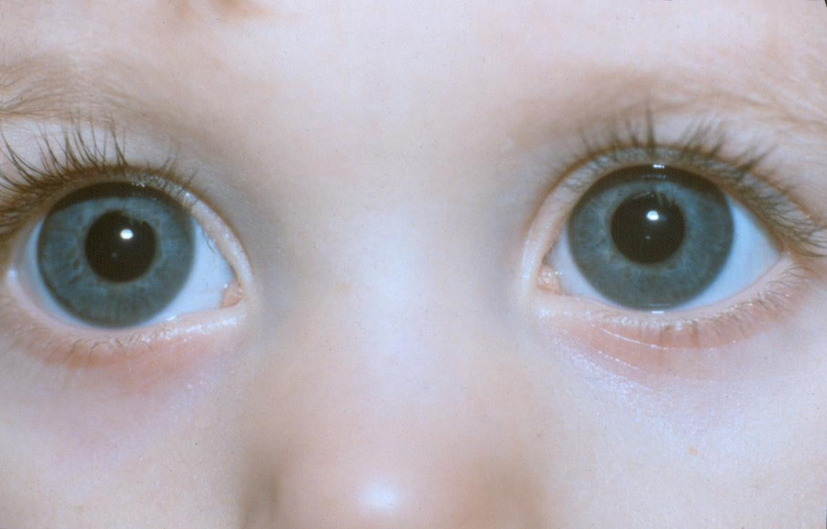
- Mata mengarah ke sudut bawah, disebut dengan “sun-setting”
- Rewel
- Kejang
- Mengantuk
- Kepala yang sangat besar atau kepala tumbuh sangat cepat dibandingkan bagian tubuh lainnya
- Muntah
Diagnosis
Diagnosis dapat dilakukan ketika bayi masih dalam kandungan atau setelah bayi lahir.
Diagnosis pre-natal (sebelum lahir)
Melakukan pemeriksaan hamil dan USG rutin selama kehamilan dapat mendeteksi masalah perkembangan otak bayi, seperti pembesaran ventrikel. Dengan teknologi pencitraan yang canggih, hidrosefalus kongenital dapat dideteksi sejak bulan ketiga atau keempat kehamilan. Pada bulan kelima atau keenam, pelebaran tidak normal pada rongga otak akan lebih jelas terlihat. Tes untuk menilai kondisi bayi sebelum lahir meliputi:
- Amniosentesis, yaitu pengambilan sampel cairan ketuban dari dalam rahim. Tes ini dapat memeriksa kromosom bayi dan mendeteksi adanya cacat lahir lain yang terkait dengan hidrosefalus.
- USG. Pemeriksaan ini dilakukan oleh dokter spesialis radiologi atau kandungan. Tes ini akan menentukan apakah ada penumpukan cairan yang tidak normal. Namun, tes ini tidak dapat menunjukkan adanya obstruksi (sumbatan). Jika ditemukan masalah pada USG, tes lanjutan dapat membantu mendiagnosis masalah dengan lebih detail.
Diagnosis post-natal (setelah lahir)
Hidrosefalus kongenital dapat dideteksi sebelum lahir, namun lebih sering didiagnosis saat lahir atau segera setelahnya. Untuk membuat diagnosis, dokter mengevaluasi kondisi fisik bayi secara menyeluruh. Dokter juga akan menanyakan riwayat kesehatan keluarga, terutama jika ada kerabat yang memiliki cacat tabung saraf. Dokter juga dapat melakukan pemeriksaan penunjang dengan teknik pencitraan seperti USG, CT scan, MRI, atau teknik pemantauan tekanan otak.
Tata Laksana
Meskipun dokter dapat mendiagnosis hidrosefalus sejak dalam kandungan, pengobatan umumnya belum dimulai sampai bayi lahir.
Penanganan yang paling umum untuk hidrosefalus kongenital adalah membuat sistem shunt. Dokter bedah akan menempatkan tabung plastik fleksibel pada otak bayi untuk mengalirkan kelebihan cairan. Ujung tabung yang lain akan dihubungkan ke perut atau tempat lain pada tubuh yang dapat menyerap kelebihan cairan serebrospinal.
Setelah pemasangan sistem shunt, pasien harus dikontrol dengan cermat. Pasien mungkin membutuhkan lebih dari satu prosedur. Masalah yang mungkin muncul setelah pemasangan shunt antara lain:
- Infeksi
- Saluran tersumbat
- Masalah mekanik
- Shunt kurang panjang
Penanganan lainnya adalah ETV (endoscopic third ventriculostomy). Prosedur ini menggunakan teknologi serat optik. Dokter akan mengarahkan kamera kecil ke otak bayi dan membuat lubang di ventrikel menggunakan alat khusus sebagai jalan keluar cairan CSF. Cairan otak kemudian mengalir melalui lubang itu dan diserap ke dalam aliran darah.
ETV juga memiliki beberapa risiko seperti lubang di ventrikel dapat tiba-tiba menutup. Hal ini dapat mengancam jiwa. Infeksi, demam, atau pendarahan juga mungkin terjadi.
Bahkan setelah dilakukan penanganan, hidrosefalus kongenital dapat memengaruhi perkembangan fisik dan intelektual. Anak mungkin memerlukan rehabilitasi dan pendidikan khusus. Namun, tetap ada harapan bagi anak untuk bisa menjalani kehidupan yang normal dengan hanya tersisa sedikit keterbatasan.
Tanyakan kepada dokter, perawat, atau terapis mengenai penanganan dan perawatan yang akan diberikan kepada anak Anda. Jika perawatan termasuk obat-obatan, pastikan anak meminumnya sesuai aturan dokter. Catat jadwal kontrol rutin agar perkembangan anak dapat terpantau dengan baik.
Komplikasi
Pada kebanyakan kasus, hidrosefalus yang tidak diobati dapat berkembang menjadi komplikasi seperti cacat intelektual, perkembangan, maupun fisik seperti gangguan belajar, gangguan bicara, gangguan memori, gangguan kemampuan sosial, gangguan penglihatan seperti juling dan penurunan tajam penglihatan, gangguan koordinasi fisik, atau epilepsi. Hidrosefalus yang berat juga dapat mengancam jiwa. Kasus yang tidak terlalu parah, bila ditangani dengan tepat, dapat memiliki sedikit komplikasi.
Pencegahan
Belum ditemukan cara untuk mencegah hidrosefalus kongenital, namun risikonya dapat dikurangi. Pastikan ibu hamil mengonsumsi makanan bergizi seimbang yang penting untuk perkembangan otak janin, serta makan makanan yang matang untuk mencegah infeksi dari makanan. Infeksi virus lain juga harus dicegah dengan menerapkan pola hidup bersih dan sehat.
Pastikan ibu melakukan pemeriksaan hamil rutin, termasuk pemeriksaan USG. Hal ini akan mengurangi risiko bayi lahir prematur, di mana bayi prematur lebih berisiko mengalami hidrosefalus. Selain itu, pemeriksaan kehamilan rutin dapat mendeteksi lebih awal jika ada masalah perkembangan pada bayi. Dengan begitu, penanganan yang tepat dapat segera dipersiapkan sebelum bayi lahir. Hal ini akan memperingan gejala dan perburukan pada perkembangan bayi.
Kapan harus ke dokter?
Segera bawa ke unit gawat darurat jika bayi mengalami tanda dan gejala berikut:
- Menangis dengan nada tinggi
- Gangguan menghisap atau gangguan makan
- Muntah berulang tanpa sebab yang jelas
- Kejang
Mau tahu informasi seputar penyakit lainnya? Cek di sini, ya!
- dr Nadia Opmalina
What is congenital hydrocephalus?. (2021). Retrieved 13 Desember 2021, from https://www.webmd.com/baby/congenital-hydrocephalus
Congenital hydrocephalus. (2021). Retrieved 13 Desember 2021, from https://www.ucsfbenioffchildrens.org/conditions/congenital-hydrocephalus
Hydrocephalus. (2021). Retrieved 30 November 2021, from https://www.mayoclinic.org/diseases-conditions/hydrocephalus/symptoms-causes/syc-20373604
Nelson SL. (2018). Hydrocephalus. Retrieved 5 Desember 2021, from https://emedicine.medscape.com/article/1135286-overview
Hydrocephalus. (2020). Retrieved 5 Desember 2021, from https://www.nhs.uk/conditions/hydrocephalus/
Hydrocephalus fact sheet. (2020). Retrieved 5 Desember 2021, from https://www.ninds.nih.gov/Disorders/Patient-Caregiver-Education/Fact-Sheets/Hydrocephalus-Fact-Sheet


