Definisi
Perdarahan retina merupakan perdarahan yang terjadi pada lapisan terdalam mata, yaitu retina, akibat pecahnya pembuluh darah. Perdarahan retina merupakan kondisi yang sering menyebabkan pasien berkunjung ke dokter mata. Retina merupakan lapisan tipis pada bagian terdalam bola mata yang sensitif terhadap cahaya karena tersusun dari sel-sel saraf. Retina mengirimkan informasi ke otak melalui saraf mata, sehingga Anda dapat melihat dan memproses informasi visual (yang terlihat). Pengobatan terhadap perdarahan retina berfungsi untuk memperlambat atau menghentikan perdarahan tersebut. Jika tidak ditata laksana dengan baik, perdarahan retina dapat menyebabkan kebutaan permanen. Perdarahan retina umumnya merupakan tanda yang membantu diagnosis dari penyakit pembuluh darah lainnya. Perdarahan retina dapat berupa titik-titik perdarahan hingga perdarahan yang banyak.
Perdarahan retina pada orang dewasa ditemukan pada usia di atas 40 tahun dengan penyakit sistemik, seperti diabetes dan hipertensi. Pada anak, perdarahan retina berhubungan dengan infeksi sistemik (menyebar ke seluruh tubuh), trauma otak, dan kelainan pembuluh darah. Pada anak baru lahir yang mengalami perdarahan retina umumnya lahir secara normal (25%) dan lahir melalui pembedahan sesar atau tindakan lain (40-50%). Perdarahan retina juga dapat terjadi pada anak yang mengalami kekerasan dalam rumah tangga, umumnya berusia kurang dari 6 bulan.
Penyebab
Perdarahan retina dapat disebabkan oleh hal-hal berikut, antara lain:
- Penyakit bola mata, seperti age-related macular degeneration (ARMD).
- Retinopati diabetikum.
- Retinopati hipertensi.
- Oklusi vena retina, di mana terjadi perdarahan retina pada seluruh bagian retina.
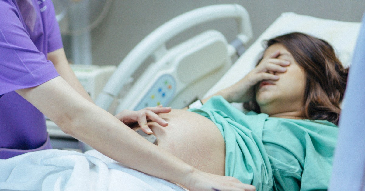
- Trauma, dapat terjadi akibat manuver valsava atau shaken baby syndrome. Trauma jalan lahir juga dapat menyebabkan perdarahan retina.
- Anemia.
- Leukemia.
- Endokarditis bakterial akut.
- Sickle cell anemia.
- Preeklamsia.
- Lupus.
- Tinggal di dataran tinggi (high altitude retinopathy).
Faktor Risiko
- Usia tua.
- Merokok.
- Obesitas.
- Memiliki diabetes, hipertensi atau penyakit sistemik lainnya.
- Trauma mata.
- Riwayat keluarga dengan penyakit retina.
Gejala
Gejala yang dapat dialami, antara lain adalah:
- Melihat bayangan hitam atau titik-titik melayang.
- Pandangan buram.
- Kerusakan pada satu sisi lapang pandang.
- Kehilangan penglihatan.
Diagnosis
Dokter Anda akan menanyakan beberapa hal, antara lain keluhan terkait dengan mata Anda, kapan Anda pertama kali merasakan keluhan tersebut, apakah keluhan tersebut dirasakan pada salah satu mata atau kedua mata, faktor risiko berkaitan dengan masalah retina, seperti merokok, memiliki kolesterol tinggi, diabetes, dan tekanan darah tidak terkontrol, serta riwayat keluarga dengan penyakit retina. Dokter Anda akan melihat kondisi retina Anda dengan oftalmoskop atau slit lamp, dengan sebelumnya meneteskan obat mata untuk memperbesar pupil (bagian hitam pada tengah mata) Anda. Pemeriksaan lain yang dapat dilakukan untuk melihat kondisi retina Anda adalah:
- Optical coherence tomography (OCT). Pemeriksaan yang digunakan untuk mendapatkan gambaran retina Anda secara jelas, melihat kondisi makula, mencari adanya pembengkakan, dan memantau respons terhadap pengobatan.
- Fundus autofluorescene (FAF). FAF dapat digunakan untuk menentukan derajat keparahan penyakit retina dengan melihat pigmen pada sel-sel yang menyusun lapisan retina.
- Fluorescein angiography. Pemeriksaan ini digunakan untuk melihat pembuluh darah retina Anda dan dibantu dengan penyinaran khusus. Pemeriksaan ini penting untuk melihat kondisi pembuluh darah retina Anda, mencari adanya pembentukan pembuluh darah baru yang mudah pecah (neovaskularisasi), pembuluh darah yang pecah, dan perdarahan yang terjadi.
Karena kondisi perdarahan retina berhubungan dengan kondisi sistemik, dokter Anda dapat melakukan pemantauan terhadap tekanan darah, indeks massa tubuh (IMT), dan gula darah sebelum melanjutkan pemeriksaan lainnya. Beberapa pemeriksaan yang perlu dilakukan berkaitan dengan penyakit yang mendasari perdarahan retina tersebut adalah:
- Diabetes dan sindrom metabolik memerlukan pemeriksaan darah lengkap dan HbA1c untuk melihat kontrol gula darah dalam waktu 3 bulan terakhir.
- Kelainan pembekuan darah, memerlukan pemeriksaan panel pembekuan darah.
- Infeksi, memerlukan pemeriksaan ELISA, FTA-ABS/RPR.
- Penyakit imun seperti SLE (lupus) memerlukan pemeriksaan ANA, anti-ds DNA.
Seluruh pemeriksaan tersebut dilakukan sesuai indikasi dokter Anda.
Tata Laksana
Tujuan utama dari tata laksana perdarahan retina adalah untuk memperlambat dan meghentikan perdarahan retina serta menjaga fungsi retina agar tetap baik. Pada beberapa kondisi, pencegahan tidak dapat dilakukan ketika kerusakan retina akibat perdarahan sudah terlalu parah, sehingga deteksi dini dari perdarahan retina sangat penting.
Tindakan
Terdapat beberapa tata laksana untuk mengatasi perdarahan retina, antara lain:
- Laser, laser dapat 'menembak' pembuluh darah yang berdarah pada retina Anda atau pembuluh darah baru yang mungkin pecah. Laser photocoagulation umumnya digunakan pada pasien dengan retinopati diabetikum. Pengobatan dengan laser dapat menyebabkan Anda kehilangan sebagian lapang pandang Anda, terutama lapang pandang bagian tepi (perifer) dan kemampuan melihat pada malam hari. Terdapat dua jenis laser photocoagulation, yaitu fokal untuk memperbaiki robekan pada retina dan scatter untuk menghentikan perdarahan.
- Vitrektomi, yaitu prosedur untuk mengambil cairan yang ada di dalam mata (vitreus) dan menggantinya dengan cairan lain. Hal ini digunakan jika perdarahan yang terjadi mengganggu retina, yang berada di belakang vitreus. Pengobatan ini dapat dilakukan pada pasien dengan robekan retina, retinopati diabetikum, infeksi, dan trauma mata.
Jika terjadi perdarahan vitreus, dokter Anda akan menyarankan untuk istirahat total terlebih dahulu untuk mencegah perdarahan lebih lanjut. Menggunakan eye patching (pelindung mata) dapat membantu mengurangi pergerakan mata dan membantu melokalisasi perdarahan. Anda juga diminta untuk berhenti mengonsumsi obat-obatan pengencer darah untuk sementara waktu.
Edukasi
Gangguan tajam penglihatan dan kehilangan penglihatan dapat mengganggu kualitas hidup Anda. Beberapa hal ini dapat membantu Anda beradaptasi dengan kondisi ini:
- Minta dokter Anda untuk meresepkan kacamata untuk membantu penglihatan Anda.
- Ubah tampilan komputer Anda dan tambahkan bantuan audio.
- Gunakan elektronik yang dapat membantu Anda melakukan aktivitas sehari-hari, seperti menggunakan audiobooks untuk mendengarkan buku.
- Pasang lampu yang lebih terang di rumah Anda.
- Tanyakan apakah Anda masih dapat menyetir kepada dokter Anda.
- Bergabung dengan komunitas yang mengalami hal yang sama untuk mendapatkan dukungan.
Komplikasi
Bayi baru lahir dan anak yang mengalami perdarahan retina akibat cedera jalan lahir memiliki gejala yang minimal dan dapat sembuh sempurna dalam waktu 2–4 minggu. Perdarahan retina yang disebabkan oleh kondisi metabolik atau oklusi vena dapat dicegah dengan mengontrol faktor risiko yang menyebabkan penyakit tersebut. Perdarahan retina yang terjadi pada daerah makula (tempat jatuhnya bayangan) dapat memiliki gejala dan komplikasi yang buruk, namun jika ditata laksana dengan cepat memiliki kemungkinan untuk kembali normal. Umumnya perdarahan retina dapat sembuh dalam waktu 3 bulan pada orang dewasa.
Komplikasi yang dapat terjadi jika perdarahan retina tidak ditangani dengan cepat, antara lain gluakoma, perluasan perdarahan retina, submacular fibrosis, neovaskularisasi retina (pembentukan pembuluh darah retina baru yang berpotensi untuk pecah di kemudian hari dan menyebabkan perdarahan retina berulang), dan kebutaan permanen.
Pencegahan
Untuk mencegah perdarahan retina, perlu dilakukan hal-hal berikut:
- Mengontrol faktor risiko yang dapat menyebabkan perdarahan retina seperti hipertensi dan diabetes.
- Menggunakan pelindung mata saat bekerja atau beraktivitas yang memiliki risiko cedera mata.
Kapan Harus ke Dokter?
Penting untuk selalu memeriksakan diri ke dokter setiap mengalami keluhan atau perubahan pada penglihatan Anda. Jika Anda melihat adanya bayangan hitam yang melayang dan penurunan penglihatan, segera periksakan diri Anda ke fasilitas kesehatan terdekat.
Sebelum ke dokter, Anda dapat mencatat seluruh keluhan penglihatan yang Anda alami serta obat-obatan yang diresepkan pada anda dan dikonsumsi rutin. Jika Anda mengalami kesulitan melihat, minta keluarga Anda untuk menemani Anda ke fasilitas kesehatan terdekat.
Ingin tahu informasi seputar penyakit lainnya? Yuk simak informasinya di sini ya!
- dr Nadia Opmalina
Retinal diseases. (2020). Mayoclinic. Available from: https://www.mayoclinic.org/diseases-conditions/retinal-diseases/diagnosis-treatment/drc-20355827
Kanukollu VM, Ahmad SS. Retinal Hemorrhage. [Updated 2021 Aug 11]. In: StatPearls [Internet]. Treasure Island (FL): StatPearls Publishing; 2021 Jan-. Available from: https://www.ncbi.nlm.nih.gov/books/NBK560777/
Jinagal J. (2019)/ Retinal hemorrhage from Blunt Ocular Trauma. N Engl J Med 2019; 381:2252.