Definition
Bronchiectasis is a long-term respiratory disorder characterized by widening the airways, leading to mucus buildup. The accumulation of mucus makes the lungs more vulnerable to infections. Bronchiectasis can occur at any age, especially in developing countries, where the availability of antibiotics is not uniform.
Causes
The respiratory system consists of small tubes called bronchi. Air flows into the lungs through the bronchi and then reaches small sacs called alveoli. In the alveoli, oxygen is absorbed into the blood, while carbon dioxide is expelled. Air flows out through the bronchi again. The inner walls of the bronchi are lined with mucus, which acts as a protective barrier against small particles trying to enter the lungs.
In bronchiectasis, one or more bronchi widens beyond the normal limits. This also means there is a buildup of mucus in the bronchi, which can make them more susceptible to infection. If an infection occurs, the bronchi become damaged, accumulating more mucus, and infections become more likely. This cycle can lead to a long-term deterioration in lung function.
Additionally, falls from significant heights, exceeding 8-10 feet, and participation in contact sports like football, rugby, or hockey may precipitate flail chest. Certain predisposing factors, including specific bone diseases, age-related bone degradation, or select cancer types, heighten susceptibility to this condition.
Rib fractures engendered by blunt trauma often manifest excruciating pain and can provoke additional injuries, such as punctured lungs or compromised blood vessels due to bone fractures. Consequently, flail chest is regarded as the most severe consequence of blunt trauma to the chest wall.
Risk factor
Bronchiectasis is caused by various factors, which can be divided into:
- Infections: Lung infections that can lead to bronchiectasis can be caused by bacteria, viruses, and fungi. Bacteria that can cause lung infections leading to bronchiectasis include Mycobacterium tuberculosis, Haemophilus influenzae, Pseudomonas aeruginosa, Staphylococcus aureus, Mycoplasma, and others. Virus infections typically associated with bronchiectasis include respiratory syncytial virus (RSV) and measles virus.
- Genetic conditions: Genetic conditions that can lead to bronchiectasis include cystic fibrosis, Young's syndrome, hypogammaglobulinemia (low levels of antibodies in the body), alpha 1-antitrypsin deficiency, Kartagener syndrome, and Mounier-Kuhn syndrome.
- Acquired conditions: Blockages in the airways due to foreign objects, mucus buildup, tumors, and enlarged lymph nodes. Diseases like gastroesophageal reflux disorder (GERD), as well as injuries to the airways, such as burns, can also cause this condition.
- Inflammation: Bronchiectasis can also be associated with diseases that cause inflammation in specific parts of the body, such as ulcerative colitis (which typically affects the large intestine), rheumatoid arthritis (which typically affects small joints like those in the fingers), and Sjögren's syndrome (which typically affects the tear and salivary glands). Other respiratory disorders like asthma, bronchomalacia, chronic obstructive pulmonary disease (COPD), and lung calcification/fibrosis can also cause this condition.
- Idiopathic (cause unknown).
Symptoms
Bronchiectasis is typically characterized by:
- Persistent productive cough, lasting for months to years
- Coughing up blood may also occur due to bronchial damage
- Shortness of breath
- Chest pain
- Wheezing
- Fever
- Fatigue
- Weight loss
Usually, bronchiectasis is accompanied by recurrent lung infections requiring continuous antibiotic therapy or a severe single infection in childhood. Childhood infections that can progress to bronchiectasis include tuberculosis, pertussis (whooping cough), and pneumonia.
Diagnosis
Diagnosis of bronchiectasis typically involves obtaining a person's medical history and conducting several examinations. These examinations can include a direct physical examination, laboratory tests, or imaging. You will undergo a chest examination aimed at identifying respiratory problems, such as additional breath sounds. Additionally, clubbing of the fingers may be found as a result of long-term oxygen deficiency.
Laboratory tests that can be performed include sputum examination, for both tuberculosis testing (with acid-fast bacilli, AFB, or rapid molecular testing, RMT) and other bacterial examinations. Sputum can also be cultured to identify the types of bacteria or fungi present. Complete blood count, levels of immune cells, substances in the body such as alpha 1-antitrypsin, and Aspergillus fungus examination can also be performed. Autoantibody tests (immune cells that attack one's cells) can also be conducted.
Meanwhile, imaging examinations that can be performed include chest X-rays, computed tomography scans (CT scans), and bronchoscopy (inserting a tube with a camera into the respiratory tract) depending on availability and disease considerations. Pulmonary function tests with spirometry can also be performed. In pulmonary function testing, you will be asked to breathe normally, and medications may be administered to identify any abnormalities in your respiratory tract.
Management
Bronchiectasis is a manageable condition, but it's rarely curable. The goal of bronchiectasis management is to identify and treat the underlying causes, improve mucus clearance in the bronchi, control infections, and open up airway blockages. To identify and treat the underlying causes, you may be referred to a more comprehensive healthcare facility if your condition cannot be managed with the existing facilities.
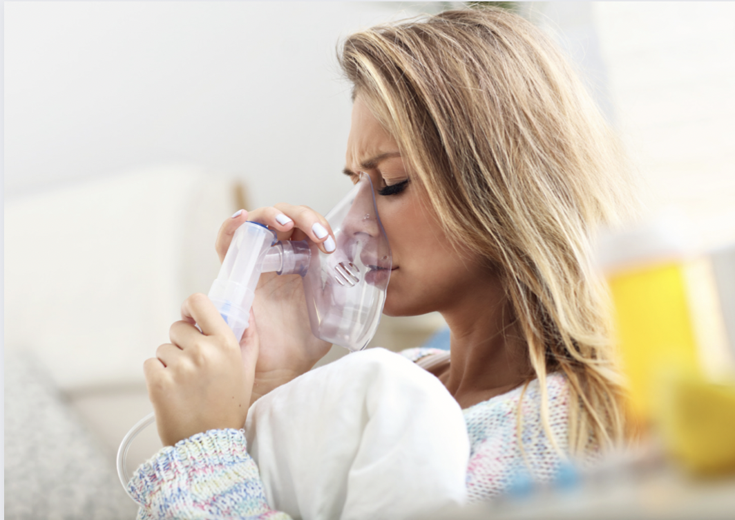
Next, mucus clearance in the bronchi can be achieved in various ways. You may be trained to perform chest percussion and vibration to help clear mucus. You may also be provided with specialized equipment to assist in mucus clearance, such as a nebulizer with a 7% sodium chloride solution.
Infection control can be done in collaboration with your doctor. If you or your child are known to have bronchiectasis, you may need regular check-ups as advised by your doctor. You may be prescribed antibiotics to prevent specific bacterial infections. You may also undergo sputum culture to guide antibiotic therapy effectively. Meanwhile, opening up airway blockages can be aided by medications similar to asthma medications.
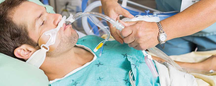
You can also make lifestyle changes, such as quitting smoking or avoiding active smokers, improving nutrition with the help of food supplements, receiving influenza and pneumococcal vaccinations (bacteria that cause pneumonia), and ensuring measles and pertussis (whooping cough) immunization status. You may also receive oxygen therapy if your condition is severe and accompanied by other dangerous conditions.
Complications
Complications of bronchiectasis are rare but typically severe. One of the most severe complications is massive hemoptysis, resulting from the rupture of blood vessels near the airways. Such hemoptysis can be life-threatening and often requires emergency intervention. Other complications may include recurrent lung infections, respiratory failure, heart disease due to lung impairment, and lung perforation (pneumothorax).
Prevention
Preventing bronchiectasis can be achieved by preventing lung infections, which are generally the main cause. One preventive measure is to ensure complete childhood immunizations, especially for measles and pertussis. You also need to supervise your children, especially young ones, to prevent them from swallowing small objects. If you suspect your child has swallowed a foreign object, take them to the nearest emergency department so the object can be removed promptly. Additionally, prevention can be achieved by not smoking and avoiding smoke or other toxic gasses. If you work in a high-risk environment for toxic gasses, always use personal protective equipment properly and completely.
When to see a doctor?
If you, your child, or someone around you has a persistent productive cough, visit the nearest doctor. This cough may not necessarily be caused by bronchiectasis but often requires further examination. If the doctor suspects bronchiectasis, you may be referred to a pulmonary or internal medicine specialist for further evaluation, depending on availability.
- dr Nadia Opmalina
Bird, K., & Memon, J. (2021). Bronchiectasis. Retrieved 16 February 2022, from https://www.ncbi.nlm.nih.gov/books/NBK430810/
Bronchiectasis. (2021). Retrieved 16 February 2022, from https://www.nhs.uk/conditions/bronchiectasis/
Bronchiectasis | NHLBI, NIH. (2020). Retrieved 16 February 2022, from https://www.nhlbi.nih.gov/health-topics/bronchiectasis
Emmons, E. (2020). Bronchiectasis: Practice Essentials, Background, Pathophysiology. Retrieved 16 February 2022, from https://emedicine.medscape.com/article/296961-overview