Definisi
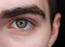
Iridodialisis adalah adanya robekan pada dasar selaput pelangi mata (iris). Hal ini umumnya disebabkan oleh cedera atau benturan langsung pada mata, benturan pada kepala, dan kecelakaan akibat benda tajam pada bola mata.
Iris merupakan bagian bola mata yang memberikan warna pada mata. Iris berfungsi untuk melebarkan maupun menyempitkan pupil. Hal ini berfungsi untuk mengatur sedikit atau banyaknya cahaya yang masuk ke dalam bola mata. Pupil akan melebar pada ruangan yang gelap untuk memasukkan lebih banyak cahaya ke dalam mata dan akan menyempit pada ruangan yang terang agar tidak terlalu banyak cahaya yang masuk ke dalam mata.
Penyebab
Penyebab dari iridodialisis adalah adanya benturan, baik langsung maupun tidak langsung pada mata, yang menyebabkan iris terlepas dari badan silia. Hal ini menyebabkan adanya dua tempat masuknya cahaya, pupil dan melalui robekan tersebut. Iridodialisis berbeda dengan siklodialisis, di mana robekan terjadi bukan pada iris, namun pada badan silia yang terletak di sisi dalam bola mata. Robekan juga dapat terjadi saat sedang melakukan operasi mata, yang disebut iatrogenik.
Karena ada dua akses cahaya (dua pupil) untuk masuk ke dalam mata, orang yang mengalami iridodialisis dapat mengalami sensasi bayangan ganda dan silau.
Faktor Risiko
Faktor risiko dari iridodialisis adalah cedera langsung pada mata atau cedera kepala.
Gejala
Gejala yang dapat terjadi pada iridodialisis antara lain:
- Tidak ada gejala pada iridodialisis yang kecil
- Pandangan ganda
- Nyeri dan silau ketika melihat cahaya
- Kilatan cahaya
- Adanya darah pada bilik mata depan (hifema)
Pada trauma mata, gejala yang dapat dialami antara lain adalah:
- Nyeri pada mata
- Pandangan buram
- Riwayat cedera kepala atau cedera bola mata
Diagnosis
Dokter Anda menyimpulkan diagnosis iridodialisis berdasarkan temuan klinis. Dokter Anda dapat melihat adanya robekan pada iris melalui pemeriksaan oftalmoskopi atau slit lamp. Selain itu, dokter Anda juga akan mencari kelainan lain pada bola mata yang disebabkan oleh benturan, seperti dislokasi lensa, hifema, ruptur bola mata, dan peningkatan tekanan bola mata.
Terdapat beberapa kondisi lain yang dapat menyerupai iridodialisis, antara lain:
- Ruptur bola mata
- Tertinggalnya benda asing pada bola mata
- Dislokasi lensa
- Iritis traumatik
- Ulkus kornea
- Endoftalmitis
Tata laksana
Tata laksana terhadap iridodialisis bergantung pada luas robekan iris tersebut. Iridodialisis yang kecil tidak memerlukan pengobatan khusus. Dokter Anda akan memberikan tata laksana berdasarkan gejala lain yang ada, seperti adanya darah pada bilik mata depan akan ditata laksana dengan manajemen hifema, sedangkan peningkatan tekanan bola mata akan ditata laksana dengan manajemen glaukoma. Jika diperlukan, dokter Anda akan melakukan pemeriksaan CT scan dan ultrasonography (USG) untuk menyingkirkan diagnosis lain, seperti adanya benda asing pada mata, ablasio retina, dislokasi lensa, dan perdarahan vitreus.
Secara umum, pilihan tata laksana pada iridodialisis, antara lain:
- Bed rest dan pemantauan perburukan gejala.
- Menggunakan kacamata untuk meredakan gejala.
- Operasi dapat dilakukan pada iridodialisis yang luas dan menyebabkan pandangan ganda. Teknik operasi yang digunakan pada iridodialisis mirip dengan operasi katarak. Dokter mata Anda akan melakukan sayatan kecil pada mata dan memasukkan benang untuk menjahit iris anda ke posisi semula. Kemudian, dokter Anda akan menjahit kembali sayatan awal tersebut. Teknik yang dilakukan akan berbeda bergantung pada kondisi iris.
Komplikasi
Komplikasi dari iridodialisis adalah glaukoma atau peningkatan tekanan bola mata. Peningkatan tekanan bola mata ini terjadi secara perlahan dan terus menerus (kronik) akibat adanya gangguan aliran cairan di dalam mata. Adanya cedera atau robekan tersebut menyebabkan terbentuknya jaringan parut yang menganggu aliran. Jika terus dibiarkan, tekanan di dalam bola mata akan semakin tinggi dan mengganggu saraf mata. Kerusakan saraf mata secara terus menerus dapat menyebabkan gangguan lapang pandang dan kebutaan.
Pencegahan
Karena penyebab dan faktor risiko dari iriodidalisis berhubungan dengan cedera pada mata maka pencegahan utama adalah mencegah terjadinya cedera pada mata. Beberapa langkah dapat dilakukan untuk mencegah cedera mata, antara lain:
- Gunakan pelindung mata ketika melakukan aktivitas yang berisiko tinggi untuk terpapar terhadap debu, benda asing, atau benda yang melayang. Perhatikan anak Anda jika sedang melakukan aktivitas berisiko tersebut.
- Gunakan pelindung mata ketika sedang memotong rumput dan jauhkan anak Anda ketika Anda sedang melakukan hal tersebut.
- Letakkan benda tajam di tempatnya dan jauhkan dari jangkauan anak-anak.
- Jangan menggunakan benda-benda yang dapat membuat jatuh terpeleset, seperti karpet yang licin. Jika Anda tinggal dengan lansia atau anak-anak, pastikan selalu berhati-hati agar tidak terjatuh dan gunakan pelindung pada sudut furnitur yang tajam agar tidak melukai ketika tidak sengaja terbentur.
- Gunakan car seat untuk anak Anda yang dilengkapi dengan sabuk pengaman yang aman. Jangan letakkan anak di bawah 12 tahun pada kursi depan. Simpan benda-benda yang dapat bergerak atau jatuh di bagasi mobil Anda.
- Berhati-hati lah dengan mainan anak Anda. Jangan berikan permainan yang berisiko seperti tembak-tembakan, pistol mainan, dan panahan. Hindari memberikan permainan seperti laser karena dapat merusak retina.
- Gunakan pelindung mata ketika sedang melakukan olahraga yang melibatkan bola, stik, raket, benda yang melayang (shuttle cock, boal tenis), dan lain-lain. Gunakan pelindung mata yang disetujui dan dilabeli ASTM F803. Pelindung mata ini telah memenuhi syarat ASTM internasional. Pelindung mata yang tidak terbukti dapat melindungi mata Anda dari cedera dalam olahraga adalah kaca mata. Atlet profesional perlu menggunakan lensa polikarbonat dengan ketebalan 2 mm untuk olahraga low impact dan 3 mm untuk olahraga high impact. Pelindung wajah seperti helm harus digunakan pada permainan hockey, sepak bola, baseball, dan lacrosse.
- Berhati-hati lah ketika membuka botol, karena tutup botol dapat melayang ke wajah Anda.
Kapan Harus ke Dokter?
Jika Anda mengalami silau dan pandangan ganda setelah benturan langsung pada bola mata, cedera kepala, terkena benda tajam pada bola mata, segera periksakan diri Anda ke fasilitas kesehatan terdekat. Walaupun cedera mata yang Anda alami terkesan ringan, penting untuk segera memeriksakan diri ke dokter agar tidak ada hal-hal lain yang dapat berdampak buruk jika tidak ditangani dengan cepat.
Sebelum ke dokter, perhatikan beberapa hal berikut:
- Jangan menyentuh, mengucek, atau menekan mata.
- Jangan teteskan obat mata apapun pada mata.
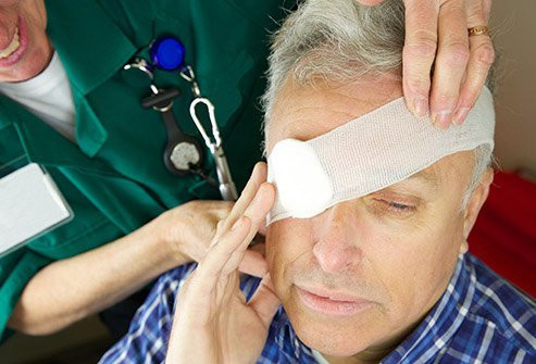
- Lindungi mata dengan menggunakan patch (penutup mata) yang tidak menempel langsung pada mata hingga Anda sampai ke fasilitas kesehatan.
- Minta orang lain untuk membantu Anda ke fasilitas kesehatan.
Mau tahu informasi seputar penyakit lainnya? Cek di sini, ya!
- dr Nadia Opmalina
Holtz CM. (2019). Iridodialysys. WikiEM. Retrieved from: https://wikem.org/wiki/Iridodialysis
Knoop K.J., & Palma J.K. (2021). Iridodialysis. Knoop K.J., & Stack L.B., & Storrow A.B., & Thurman R(Eds.), The Atlas of Emergency Medicine, 5e. McGraw Hill. https://accessmedicine.mhmedical.com/content.aspx?bookid=2969§ionid=250455915
Columbia University Department of Ophtalmology. (N/A). Traumatic Iridodialysis. ColumbiaEye. Retrieved from: https://www.columbiaeye.org/education/digital-reference-of-ophthalmology/glacucoma/angle-closure-glaucoma/traumatic-iridodialysis
Columbia University Department of Ophtalmology. (N/A). Iridodialysis. ColumbiaEye. Retrieved from: https://www.columbiaeye.org/education/digital-reference-of-ophthalmology/cornea-external-diseases/trauma/iridodialysis
Luviano D. (N/A). Traumatic iridodialysis. AAO. Retrieved from: https://www.aao.org/image/traumatic-iridodialysis-4
MayoClinic Staff. (2019). Eye injury: Tips to protect vision. MayoClinic. Available from: https://www.mayoclinic.org/healthy-lifestyle/adult-health/in-depth/eye-injury/art-20047121


