")

")
Definisi
Obstruksi duktus nasolakrimal adalah istilah untuk keadaan di mana terjadi penyumbatan pada saluran air mata. Kondisi ini merupakan kelainan sistem kelenjar air mata yang paling sering terjadi.
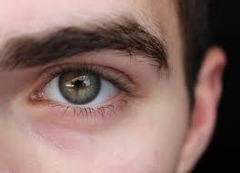
Air mata diproduksi oleh kelenjar lakrimal atau kelenjar air mata. Kelenjar ini terletak di dalam kelopak mata atas. Normalnya, air mata mengalir dari kelenjar lakrimal ke lubang (puncta) di sudut dalam kelopak mata atas dan bawah. Puncta mengarah ke kanal kecil yang akan mengalirkan air mata ke kantung air mata di sisi hidung (kantung lakrimal). Dari sana air mata mengalir ke arah hidung melalui saluran (duktus nasolakrimalis). Setelah sampai di hidung, air mata akan diserap kembali.
Penyumbatan dapat terjadi pada setiap titik dalam sistem drainase air mata, mulai dari puncta hingga ke hidung. Ketika hal itu terjadi, air mata tidak dapat mengalir dengan baik sehingga membuat mata berair dan meningkatkan risiko peradangan serta infeksi pada mata.
Penyumbatan saluran air mata sering terjadi pada bayi baru lahir. Sekitar 6-20% bayi baru lahir memiliki gejala sumbatan saluran air mata. Keluhan biasanya muncul pada minggu hingga bulan pertama kehidupan, ditandai dengan keluarnya air mata dan kotoran mata secara berlebihan. Kondisi ini biasanya akan membaik tanpa pengobatan pada tahun pertama kehidupan.
Pada orang dewasa, saluran air mata yang tersumbat mungkin disebabkan oleh cedera, infeksi, atau tumor. Saluran air mata yang tersumbat hampir selalu dapat diperbaiki. Terapi yang dipilih tergantung pada penyebab penyumbatan dan usia orang yang terkena.
Penyebab
Sumbatan saluran air mata dapat terjadi pada semua usia, mulai dari bayi baru lahir hingga dewasa. Penyebabnya antara lain:
- Kelainan bawaan. Banyak bayi lahir dengan saluran air mata yang tersumbat. Sistem drainase air mata bayi tersebut mungkin tidak sepenuhnya berkembang atau ada kelainan saluran. Seringkali juga terdapat selaput tipis yang menutupi lubang saluran yang mengarah ke hidung.
- Pertambahan usia. Seiring bertambahnya usia, puncta dapat menyempit dan menyebabkan penyumbatan.
- Infeksi atau peradangan. Infeksi kronis atau peradangan pada mata, gangguan sistem drainase air mata atau hidung dapat menyebabkan saluran air mata tersumbat.
- Cedera atau trauma. Cedera pada wajah dapat menyebabkan kerusakan tulang atau jaringan parut di dekat sistem drainase sehingga mengganggu aliran air mata. Bahkan, partikel kecil dari kotoran atau sel kulit yang lepas jika masuk ke saluran dapat menyebabkan penyumbatan.
- Tumor. Tumor di hidung atau bagian lain dari sistem drainase air mata dapat menyebabkan penyumbatan.
- Obat tetes mata. Meski kasusnya jarang, namun penggunaan obat-obatan tertentu dalam jangka panjang, seperti obat tetes mata untuk glaukoma, dapat menyebabkan saluran air mata tersumbat.
- Pengobatan kanker. Saluran air mata yang tersumbat dapat timbul karena efek samping dari kemoterapi dan radiasi untuk kanker.
Faktor Risiko
Faktor-faktor tertentu dapat meningkatkan risiko Anda mengalami sumbatan saluran air mata, antara lain:
- Usia. Semakin bertambah usia, risiko mengalami sumbatan saluran air mata juga bertambah.
- Peradangan mata kronis. Jika mata Anda terus-menerus iritasi, merah, dan meradang, Anda berisiko lebih tinggi mengalami sumbatan saluran air mata.
- Riwayat operasi daerah mata. Riwayat operasi mata, kelopak mata, hidung, atau sinus dapat menimbulkan beberapa jaringan parut pada sistem saluran dan memungkinkan timbulnya sumbatan pada saluran air mata.
- Glaukoma. Pengobatan glaukoma dengan obat tetes mata biasanya digunakan dalam waktu lama. Jika Anda menggunakan obat tetes mata anti-glaukoma atau obat tetes mata lainnya dalam jangka lama, Anda berisiko lebih tinggi mengalami sumbatan saluran air mata.
- Riwayat pengobatan kanker. Jika Anda pernah menjalani radiasi atau kemoterapi untuk mengobati kanker, terutama jika radiasi difokuskan pada wajah atau kepala, Anda berisiko lebih tinggi mengalami penyumbatan saluran air mata.
Gejala
Tanda dan gejala dari sumbatan saluran air mata antara lain:
- Keluar air mata dan kotoran mata berlebihan
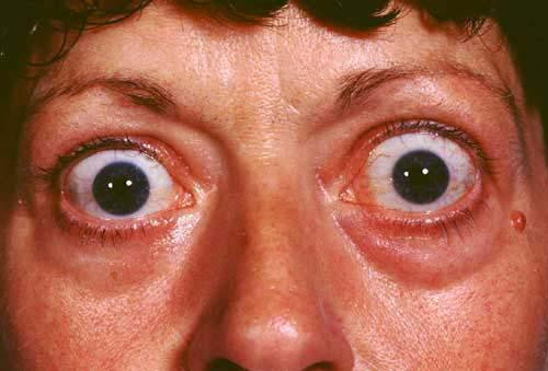
- Kemerahan pada mata
- Infeksi atau peradangan mata berulang
- Pembengkakan yang nyeri di dekat sudut dalam mata
- Krusta (kerak) pada kelopak mata
- Keluar lendir atau nanah dari kelopak dan permukaan mata
- Penglihatan kabur
Diagnosis
Untuk mendiagnosis kondisi Anda, dokter akan melakukan wawancara mengenai gejala yang Anda rasakan serta melakukan pemeriksaan fisik dan penunjang pada mata. Dokter juga akan memeriksa bagian dalam hidung untuk menentukan apakah ada gangguan struktural pada saluran hidung yang dapat menyebabkan penyumbatan. Jika dokter mencurigai adanya sumbatan saluran air mata, dokter akan meminta Anda menjalani tes tertentu untuk menemukan lokasi penyumbatan. Tes yang digunakan untuk mendiagnosis saluran air mata yang tersumbat antara lain:
- Tes drainase air mata. Tes ini mengukur seberapa cepat air mata Anda mengalir. Satu tetes pewarna khusus diletakkan pada permukaan kedua mata. Terdapat sumbatan saaluran air mata jika setelah lima menit sebagian besar pewarna masih berada di permukaan mata.
- Irigasi dan probing. Dokter akan mengalirkan larutan saline (larutan yang kandungannya mirip dengan cairan tubuh) melalui sistem drainase air mata Anda untuk memeriksa seberapa baik larutan itu mengalir. Atau dokter dapat memasukkan alat kecil (probe) ke saluran air mata melalui puncta untuk memeriksa apakah ada penyumbatan. Pada beberapa kasus, pemeriksaan ini dapat menyelesaikan masalah sumbatan yang terjadi.
- Tes pencitraan mata. Pada prosedur ini, pewarna kontras dialirkan ke sistem drainase air mata Anda melalui puncta. Kemudian X-ray, computerized tomography (CT), atau magnetic resonance imaging (MRI) digunakan untuk menemukan lokasi dan penyebab sumbatan.
Tata Laksana
Terapi yang akan Anda peroleh bergantung pada penyebab sumbatan. Anda mungkin membutuhkan lebih dari satu jenis terapi untuk memperbaiki masalah. Beberapa pilihan terapi pada sumbatan saluran air mata antara lain:
- Obat infeksi
Jika dokter mencurigai adanya infeksi, dokter akan meresepkan obat tetes mata atau pil antibiotik.
- Observasi atau pemijatan
Bayi yang lahir dengan saluran air mata yang tersumbat seringkali sembuh tanpa perawatan apa pun. Ini dapat terjadi saat sistem drainase sudah matang dalam beberapa bulan pertama kehidupan. Terkadang, ada selaput tipis yang menutupi lubang saluran yang mengarah ke hidung. Jika penyumbatan saluran air mata bayi Anda tidak membaik, dokter akan mengajarkan teknik pijat khusus untuk membantu membuka selaput tersebut.
Jika Anda mengalami cedera wajah yang menyebabkan saluran air mata tersumbat, dokter akan menyarankan Anda menunggu beberapa bulan untuk melihat apakah kondisinya membaik saat cedera Anda sembuh (mengobservasi). Saat pembengkakan akibat cedera berkurang, saluran air mata Anda akan terbuka dengan sendirinya
- Dilatasi, probing dan flushing
Untuk bayi, teknik ini dilakukan dengan bius umum. Dokter memperbesar lubang puncta dengan alat pelebar dan memasukkan probe tipis melalui puncta ke dalam sistem drainase air mata.
Untuk orang dewasa dengan puncta yang menyempit, dokter akan melebarkan puncta dengan probe kecil lalu mengirigasi saluran air mata. Ini adalah prosedur rawat jalan sederhana yang sering menjadi terapi sementara
- Pemasangan stent atau intubasi
Prosedur ini biasanya dilakukan dengan menggunakan bius umum. Sebuah pipa tipis, terbuat dari silikon atau poliuretan, dimasukkan melalui salah satu atau kedua puncta. Pipa ini kemudian melewati sistem drainase air mata ke dalam hidung Anda. Lingkaran kecil pipa akan terlihat di sudut mata Anda, dan tabung biasanya akan dibiarkan selama sekitar tiga bulan. Kemungkinan komplikasi yang mungkin terjadi adalah peradangan karena keberadaan pipa di dalam saluran
- Dilatasi kateter balon
Jika terapi lain tidak berhasil atau timbul penyumbatan lagi, prosedur ini dapat digunakan. Prosedur ini biasanya efektif untuk bayi dan balita serta untuk orang dewasa dengan penyumbatan parsial. Pertama, pasien akan diberikan bius umum. Kemudian dokter memasukkan pipa kateter ke dalam saluran air mata yang tersumbat. Terdapat balon kempis pada ujung kateter. Dokter akan mengembang dan mengempiskan balon tersebut beberapa kali untuk membuka sumbatan.
- Dakrosistorinostomi
Prosedur ini membuat jalan bagi air mata untuk mengalir keluar dari hidung dan dilakukan pemasangan stent agar saluran tetap terbuka selama proses penyembuhan. Anda akan diberi bius umum atau lokal jika dilakukan sebagai prosedur rawat jalan. Setelah operasi, Anda akan menggunakan semprotan hidung dan obat tetes mata khusus untuk mencegah infeksi dan mengurangi peradangan. Anda akan kontrol ke dokter lagi setelah 6-12 minggu untuk melepas stent .
Jika tumor yang menjadi penyebab saluran air mata Anda tersumbat, pengobatan akan difokuskan pada tumor tersebut. Pembedahan dapat dilakukan untuk mengangkat tumor atau dokter akan merekomendasikan terapi lain untuk mengecilkan tumor.
Komplikasi
Air mata yang tersisa di sistem drainase akan menggenang akibat sistem drainase mengalami gangguan. Genangan ini dapat menjadi tempat tumbuhnya bakteri, virus, atau jamur yang dapat menyebabkan infeksi dan peradangan mata berulang. Setiap bagian dari sistem drainase air mata, termasuk selaput bening di permukaan mata (konjungtiva), dapat terinfeksi atau meradang karena saluran air mata yang tersumbat.
Pencegahan
Untuk mengurangi risiko sumbatan saluran air mata, segera periksakan mata Anda jika mengalami peradangan atau infeksi. Berikut beberapa tips untuk menghindari infeksi pada mata:
- Cuci tangan secara menyeluruh dan sering.
- Menghindari menggosok mata.
- Jangan berbagi produk mata seperti obat tetes mata atau kosmetik dengan orang lain.
- Jika memakai softlens, jaga kebersihannya sesuai dengan rekomendasi yang diberikan oleh produsen dan dokter Anda.
Kapan Harus ke Dokter?
Temui dokter Anda jika Anda terus-menerus mengeluarkan air mata selama beberapa hari atau jika mata Anda berulang kali terkena infeksi. Saluran air mata yang tersumbat dapat disebabkan oleh tumor yang menekan sistem drainase air mata. Identifikasi dini tumor dapat memberi Anda lebih banyak pilihan pengobatan.
Mau tahu informasi seputar penyakit lainnya? Cek di sini, ya!
- dr Ayu Munawaroh, MKK
Worak S. (2018). Nasolacrimal Duct Obstruction and Epiphora. Medscape. Retrieved 20 November 2021, from https://emedicine.medscape.com/article/1210141-overview.
Worak S. (2018). Nasolacrimal Duct Obstruction and Epiphora. Medscape. Retrieved 20 November 2021, from https://emedicine.medscape.com/article/1210141-clinical.
Blocked tear duct. (2021). Mayo Clinic. Retrieved 20 November 2021, from https://www.mayoclinic.org/diseases-conditions/blocked-tear-duct/symptoms-causes/syc-20351369.
Perez Y, Patel BC, Mendez MD. Nasolacrimal Duct Obstruction. Ncbi.nlm.nih.gov. Retrieved 20 November 2021, from https://www.ncbi.nlm.nih.gov/books/NBK532873/.
Lueder GT. Nasolacrimal Duct Obstruction in Children. American Academy of Ophthalmology. (2015). Retrieved 20 November 2021, from https://www.aao.org/disease-review/nasolacrimal-duct-obstruction-4.
Blocked Tear Duct (Nasolacrimal Duct). (2021). Cleveland Clinic. Retrieved 20 November 2021, from https://my.clevelandclinic.org/health/diseases/17260-blocked-tear-duct-nasolacrimal-duct-obstruction.