Definition
A choledochal duct cyst is a congenital defect that causes an abnormal dilation of the bile duct. The liver produces bile fluid, which travels to the intestines through intrahepatic and extrahepatic ducts, including the hepatic duct, common bile duct, and cystic duct. Bile fluid will combine with pancreatic enzymes along its path. This occurs due to the connection between the pancreatic duct and the choledochal duct. Bile and pancreatic enzymes will be sent into the small intestine through the ampulla of Vater to aid in food digestion. There are 5 types of choledochal duct cysts, classified based on the site of the duct expansion.
- Type I is the most common type (80–90%). The common bile duct is dilated. This expansion does not create a pouch.
- Type II choledochal cyst involves a sac protruding from the choledochal duct.
- Type III choledochal cyst is characterised by the presence of a sac near the duodenum or at the junction of the choledochal duct with the pancreatic duct.
- Type IVa involves dilation of both the intrahepatic and extrahepatic ducts.
- In Type IVb, there is dilatation specifically in the extrahepatic ducts.
- Caroli's disease, also known as Type V, is characterised by the dilation of the intrahepatic duct
Choledochal duct cysts are uncommon, occurring in around 1 in 100,000–150,000 children in western countries. Women are four times more likely to develop choledochal duct cysts. This syndrome is more prevalent in countries in East Asia, particularly Japan, but the underlying reasons for this occurrence remain unknown.
Causes
The liver produces bile, a liquid that aids in the breakdown of fats. Bile typically moves from the liver through ducts to the intestines. Abnormal enlargement of the duct causes bile to flow back to the liver. This backflow can lead to hepatic inflammation. Pancreatitis can occur due to an abnormal enlargement that hinders the flow of pancreatic enzymes to the small intestine, potentially damaging the pancreas.
The primary aetiology of choledochal duct cysts remains unidentified, but it is believed to result from the failure of the bile duct and pancreatic duct to merge properly during pregnancy. The pancreas secretes enzymes crucial for food digestion. Pancreatic enzymes leak into nearby tissue when there are problems with the connection between the bile duct and the pancreatic duct. This changes the tissue around it. These modifications result in the formation of a bag that generates a choledochal duct cyst.
Risk Factors
Factors that increase the risk of developing choledochal duct cysts include:
- Female
- East Asian and Southeast Asian descent
- Family history of choledochal duct cysts
Symptoms
Indications of a choledochal duct cyst include the following:
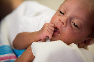
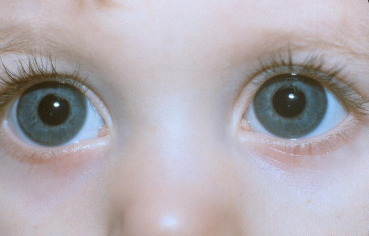
- The baby has jaundiced skin. This results from an obstructed bile flow. Neonates may exhibit jaundiced skin. This situation is typical until the baby reaches 2 weeks of age. If the yellow hue continues for over 2 weeks, it may indicate an early indication of an issue with the baby's bile.
- Intermittent abdominal pain
- A distended abdomen and a detectable mass in the stomach
- Light-coloured stools.
- Fever may arise in cases of cholangitis.
Symptoms in older infants may consist of pain, fever, nausea, vomiting, and jaundice.
Diagnosis
A clinician can diagnose the condition by reviewing the disease history, conducting a physical examination of the baby, and performing additional supporting tests. The doctor will investigate symptoms associated with jaundice in the newborn, abdominal pain, and a palpable bulge in the stomach. Approximately 85% of children with choledochal duct cysts exhibit two out of these three symptoms. Children under 12 years old may exhibit signs like jaundiced skin, light-colored faeces, and vomiting.
Following an inquiry regarding symptoms, the doctor will do various examinations, such as inspecting the baby's skin, eyes, and stomach. The doctor can analyse the poo in the nappy to verify its pale colour. On rare occasions, a mobile phone camera's pale image of the color may not accurately depict the color, necessitating a physical examination by the doctor to confirm the stool's color.
Examinations supported include:
- Haematological analysis. No blood test can conclusively diagnose a choledochal duct cyst. Additional blood panels can assist in excluding alternative diagnoses, such as infection.
- Ultrasonography (USG). An ultrasound can assist the doctor in examining the anatomical structure of the baby's biliary system. In addition to visualising the bile ducts, ultrasonography can also assist physicians in observing the blood vessels surrounding the bile ducts.
-
- Ultrasound can be used during pregnancy to diagnose a choledochal duct cyst prenatally.
- Magnetic resonance cholangiopancreatography (MRCP). This examination is more comprehensive than ultrasonography. Prior to the checkup, the infant will be administered general anaesthesia. This examination can offer a more detailed image of the bile ducts.
- CT Cholangiography. CT cholangiography enables visualisation of duct dilation in the liver and surrounding the pancreas. This examination is most suitable for type IV and V choledochal duct cysts.
- ERCP. ERCP is the most accurate diagnostic method for choledochal duct cysts. However, its invasive nature and potential consequences from radiation exposure limit its usage to instances with a high likelihood of the condition. ERCP can be used for both diagnostic and therapeutic purposes.
Management
Surgery is the treatment for choledochal duct cysts. The procedure can be conducted using open surgery or laparoscopy. Laparoscopic surgery involves a small incision in the stomach, whereas open surgery involves a bigger incision. Both of these methods may result in positive results.
The procedure often involves excising the dilated portion of the duct and extracting the gallbladder. New pathways will be established to facilitate the flow of bile from the liver to the intestines. The doctor can obtain a biopsy sample from the liver to evaluate its status.
The optimal timing for surgery in asymptomatic infants with choledochal duct cysts is still a subject of controversy. Some specialists suggest quick surgery at 6 months of age.
Although there is a risk of problems, including bleeding and infection, the majority of surgeries achieve positive outcomes. The infant can be discharged one week post-surgery. The doctor will request follow-up appointments to monitor the baby's liver function and schedule regular ultrasound examinations.
Complications
If left untreated, a choledochal duct cyst might lead to numerous complications.
- Hepatic injury (fibrosis and cirrhosis)
- Infection (Cholangitis)
- Biliary duct injury
- Rupture of a cyst
- Difficulties with food absorption and growth
- Cholelithiasis
- Pancreatitis
- Increased risk of bile duct cancer
Prevention
Prevention of choledochal duct cysts is not possible. Early identification of this disease can provide prompt care for your child to prevent complications. Over 90% of infants with choledochal duct cysts who receive surgical treatment experience positive and normal growth.
When to See a doctor?
It is important to have your infant evaluated for jaundice if he is above 2 weeks old, exhibits pale stools, a fever, and vomits.
- dr Nadia Opmalina
Children's Liver Disease Foundation. (2016). Choledocal Cyst: A Guide. Available from: https://childliverdisease.org/wp-content/uploads/2019/07/Choledochal-Cyst.pdf
Hoilat GJ, John S. Choledochal Cyst. [Updated 2021 Nov 7]. In: StatPearls [Internet]. Treasure Island (FL): StatPearls Publishing; 2022 Jan-. Available from: https://www.ncbi.nlm.nih.gov/books/NBK557762/
International Society of Ultrasound in Obstetrics and Gynecologi (ISUOG). (2019). Choledocahal cyst. Available from: https://www.isuog.org/uploads/assets/f5643e8e-e3b7-487e-bcaab5b33679bb8b/Choledochal-cyst.pdf
Henderson R. (2015). Choledochal cysts. Patientinfo. Available from: https://patient.info/doctor/choledochal-cysts
Seattle Children's GAstroenterology and Hepatology Department. Choledochal cysts. seattlechildrens. Available from: https://www.seattlechildrens.org/conditions/choledochal-cyst/
Tommolino E. (2020). Choledochal cysts. Medscape. Available from: https://emedicine.medscape.com/article/172099-overview#a1


