Definisi
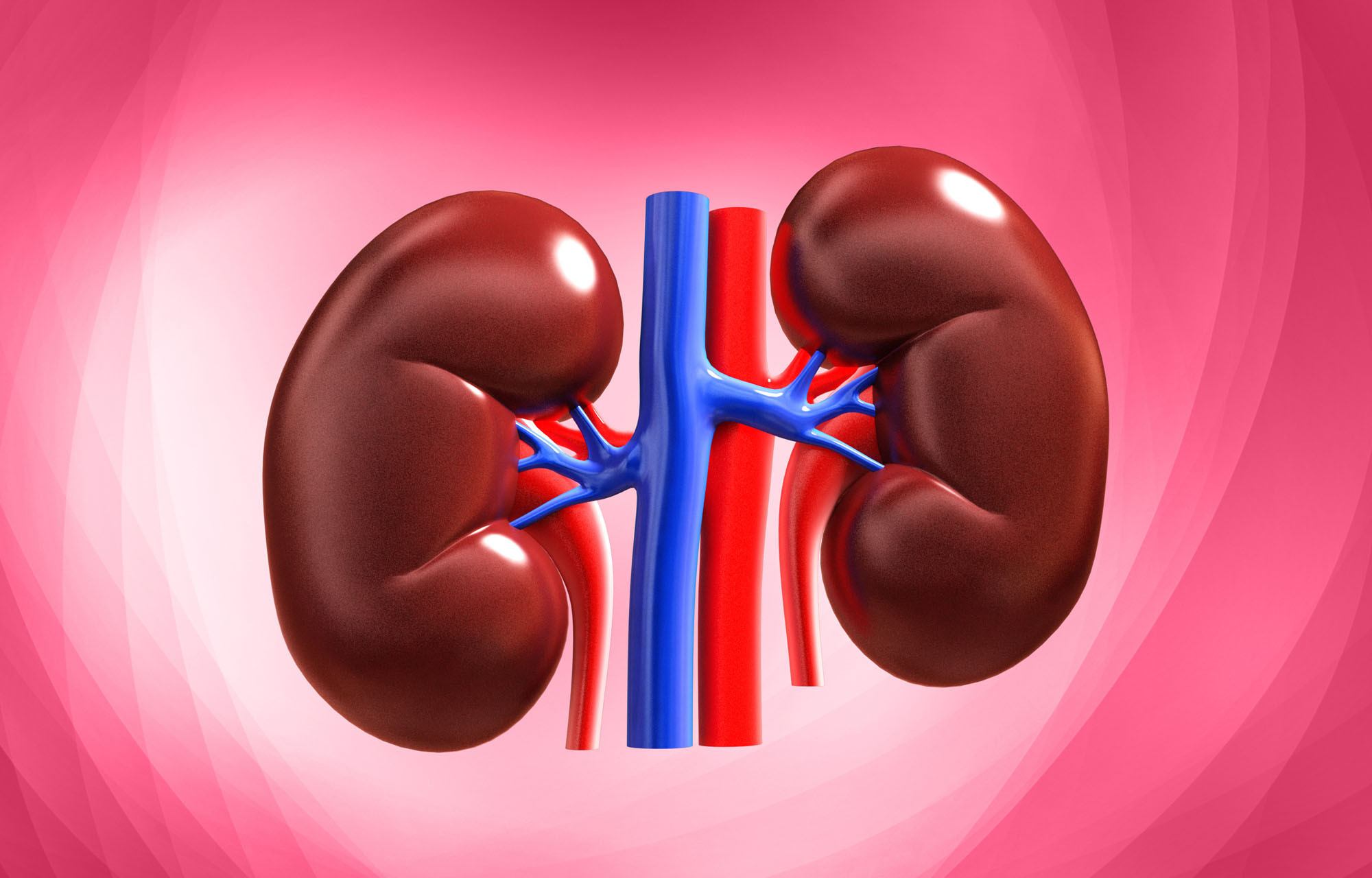
Obstruksi saluran kemih atau uropati obstruktif adalah sumbatan yang menghambat aliran urine di sepanjang saluran kemih, termasuk ginjal, ureter, kandung kemih, dan uretra. Seharusnya urine mengalir melalui ginjal ke kandung kemih, tetapi jika terjadi obstruksi, urine dapat kembali lagi ke ginjal (refluks).
Ureter memiliki 2 tabung untuk menyalurkan urine dari ginjal ke kandung kemih. Adanya uropati obstruktif dapat menyebabkan pembengkakan dan kerusakan pada salah satu atau kedua ginjal, kelainan anatomi, serta striktur (penyempitan saluran).
Kondisi ini dapat menyerang pria dan wanita dari segala usia. Obstruksi saluran kemih juga bisa menjadi masalah bagi janin selama kehamilan.
Penyebab
Penyebab uropati obstruktif yang paling sering menurut usia adalah:
- Pada anak-anak: kelainan struktur, contohnya adalah adanya katup pada bagian belakang dari uretra dan penyempitan pada ureter atau uretra
- Pada dewasa muda: batu pada ginjal, ureter, atau tempat lain di sepanjang saluran kemih
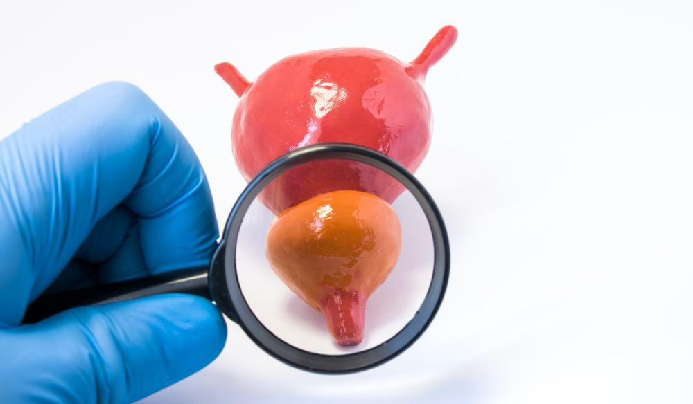
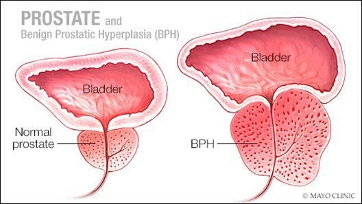
- Pada dewasa lanjut: hiperplasia prostat jinak (pembesaran prostat), kanker prostat, tumor, dan batu saluran kemih
Penyebab lain yang lebih jarang menyebabkan uropati obstruktif meliputi:
- Polip ureter (pertumbuhan jaringan)
- Gumpalan darah di ureter
- Tumor di dalam atau di dekat ureter
- Kelainan otot atau saraf pada ureter atau kandung kemih. Hal ini dapat disebabkan oleh penggunaan obat-obatan tertentu, kelainan bawaan, ataupun cedera saraf tulang belakang
- Pembentukan jaringan luka di dalam atau sekitar ureter akibat operasi, terapi radiasi, atau obat-obatan
- Adanya penonjolan pada bagian bawah atau ujung ureter ke dalam kandung kemih (uretosel)
- Tumor, abses, dan kista (benjolan berisi cairan) kandung kemih, leher rahim, rahim, prostat, atau organ panggul lainnya
- Feses yang besar dan menumpuk di rektum
Hidronefrosis atau pembengkakan pada kedua ginjal dapat terjadi saat kehamilan karena rahim yang membesar menekan ureter. Kondisi ini disebut dengan hidronefrosis kehamilan, yang biasanya sembuh sendiri setelah kehamilian berakhir, meskipun bagian ginjal dan ureter dapat tetap membengkak setelahnya.
Faktor Risiko
Terdapat beberapa faktor yang dapat meingkatkan risiko seseorang mengalami uropati obstruktif:
- Pria berusia lebih dari 60 tahun lebih mungkin terkena karena seiring pertambahan usia pria, kelenjar prostat cenderung akan bertambah besar
- Kehamilan
Gejala
Timbulnya uropati obstruktif dapat bersifat sangat cepat dan akut, atau lambat dan progresif. Anda akan merasakan sakit di bagian pinggang pada salah satu atau kedua sisi tubuh. Tingkat dan lokasi rasa sakit bervariasi pada setiap orang tergantung organ yang terlibat.
Demam, mual, dan muntah merupakan gejala umum uropati obstruktif. Anda mungkin mengalami pembengkakan atau nyeri pada pinggang (lokasi ginjal) saat urine mengalir kembali ke ginjal.
Perubahan pada kebiasaan buang air kecil dapat mengindikasikan adanya penyumbatan pada ureter. Gejalanya meliputi:
- Kesulitan buang air kecil (BAK)
- Aliran urine yang melambat
- Frekuensi buang air kecil meningkat, terutama pada malam hari
- Merasa kandung kemih tidak benar-benar kosong setelah BAK
- Penurunan jumlah keluaran (output) urine
- Terdapat darah pada urine
Uropati obstruktif juga dapat terjadi pada janin di dalam kandungan. Salah satu tanda obstruksi pada janin adalah jumlah cairan ketuban yang lebih rendah dari normal. Urine merupakan salah satu komponen di dalam cairan ketuban. Janin yang mengalami uropati obstruktif tidak dapat buang air kecil, sehingga volume cairan ketuban dapat menurun. Hal ini dapat menyebabkan komplikasi pada janin atau persalinan.
Diagnosa
Untuk menegakkan diagnosis dokter akan melakukan wawancara, pemeriksaan fisik dan pemeriksaan penunjang. Dalam wawancara dokter akan menanyakan gejala yang Anda rasakan, sejak kapan gejala timbul, obat-obatan yang sedang Anda konsumsi, serta riwayat penyakit terdahulu. Selanjutnya, dokter akan melakukan pemeriksaan fisik untuk membantu menegakkan diagnosis. Jika diperlukan, dokter juga dapat melakukan pemeriksaan penunjang, berupa:
- Tes Urine
- Pemeriksaan ini dapat melihat apakah terdapat darah pada urine atau adanya infeksi
- Pemeriksaan darah
- Jika kadar ureum dan kreatinin dalam darah abnormal, hal ini dapat menunjukkan telah terjadi gangguan pada ginjal
- Jika ditemukan jumlah sel darah putih yang meningkat, maka hal ini dapat menjadi penanda adanya infeksi
- Jika jumlah sel darah merah menurun dapat menandakan telah terjadi gangguan pada kerja ginjal
- Pemeriksaan radiologi. Pemeriksaan ini dapat dilakukan untuk mengidentifikasi bukti adanya obstruksi, seperti hidronefrosis atau lokasi sumbatan itu sendiri. Pemeriksaan ini meliputi ultrasonografi (USG), CT Scan, MRI, dan voiding cystourethrography (VCUG)
- Sistoskopi (cyctoscopy). Pemeriksaan ini dilakukan dengan memasukkan alat yang memiliki kamera untuk melihat kandung kemih dan uretra
Tata Laksana
Tata laksana biasanya bertujuan untuk menghilangkan penyebab obstruksi. Sebagai contoh, jika uretra tersumbat akibat pembesaran prostat, maka terapi akan menggunakan obat-obatan, operasi, atau pelebaran uretra dengan alat. Terapi lain seperti litotripsi atau operasi endoskopik dapat diperlukan untuk mengeluarkan batu yang menyumbat aliran urine di ureter atau ginjal.
Jika penyebab sumbatan tidak dapat ditangani dengan cepat, terutama jika terdapat infeksi, gagal ginjal, atau nyeri berat, maka akan dilakukan pengosongan saluran kemih. Ketika hidronefrosis disebabkan oleh obstruksi yang tidak mudah ditangani, maka urine yang menumpuk di atas lokasi sumbatan dapat dialirkan melalui selang yang dimasukan ke bagian belakang ginjal atau melalui selang plastik lembut yang menghubungkan kandung kemih dengan ginjal (stent ureter).
Komplikasi prosedur ini adalah pergeseran lokasi selang, infeksi, dan rasa tidak nyaman. Jika lokasi obstruksinya ada di uretra dan harus ditangani segera, maka dokter akan memasukan kateter karet lembut ke dalam kandung kemih untuk mengalirkan urine.
Obstruksi yang menyebabkan hidronefrosis kronis (jangka panjang dan perlahan) biasanya tidak memerlukan penanganan segera. Selain untuk menangani obstruksi, terapi juga bertujuan untuk menangani komplikasi.
Komplikasi
Obstruksi biasanya dapat diterapi, namun jika terlalu lama, obstruksi dapat menimbulkan beberapa komplikasi, seperti:
- Gagal ginjal
- Infeksi
- Batu lebih mungkin untuk terbentuk
Pencegahan
Anda tidak dapat sepenuhnya mencegah obstruksi saluran kemih. Namun, Anda dapat mencoba cara untuk menurunkan risiko terkena kondisi ini dengan:
- Merubah kebiasaan berkemih. Berkemih lah saat Anda merasa ingin berkemih. Sering menahan keinginan berkemih dapat melelahkan otot kandung kemih dan berisiko menyebabkan infeksi saluran kemih
- Konsumsi obat sesuai anjuran dokter. Pria dengan masalah prostat, seperti hiperplasia prostat jinak (BPH), harus mengonsumsi obat yang diresepkan oleh dokter dan menghindari obat-obatan yang dapat memicu gangguan aliran urine, seperti obat flu dan obat anti inflamasi non steroid (OAINS)
- Melakukan olahraga otot panggul atau latihan Kegel untuk menguatkan otot panggul dan meningkatkan fungsi kandung kemih dan usus
- Modifikasi diet dan gaya hidup. Obstruksi urine akibat konstipasi dapat dicegah dengan melakukan diet dan gaya hidup yang sehat. Untuk mencegah konstipasi, makanlah cukup serat, minum air yang cukup, serta melakukan aktivitas fisik rutin
Kapan Harus ke Dokter ?
Jika Anda memiliki gejala obstruksi saluran kemih, segera periksakan diri Anda ke dokter. Selain itu, jika Anda tidak dapat berkemih sama sekali atau merasakan nyeri yang hebat di area pinggang atau perut bawah, maka segera cari pertolongan medis.
Mau tahu informasi seputar penyakit lainnya? Cek di sini, ya!
- dr Nadia Opmalina
Urinary Tract Obstruction - Kidney and Urinary Tract Disorders - MSD Manual Consumer Version. (2022). Retrieved 5 July 2022, from https://www.msdmanuals.com/home/kidney-and-urinary-tract-disorders/obstruction-of-the-urinary-tract/urinary-tract-obstruction
Retention, P., & Health, N. (2022). Preventing Urinary Retention | NIDDK. Retrieved 5 July 2022, from https://www.niddk.nih.gov/health-information/urologic-diseases/urinary-retention/prevention
Urinary obstruction. (2022). Retrieved 5 July 2022, from https://www.health.govt.nz/your-health/conditions-and-treatments/diseases-and-illnesses/urinary-problems/urinary-obstruction
Erica Roth. Obstructive Uropathy. (2018). Retrieved 15 July 2022, from Urinary Tract Obstruction: Types, Causes, and Symptoms (healthline.com)
Medscape. Urinary Tract Obstruction. (2020). Retrieved 15 July 2022, from Urinary Tract Obstruction: Practice Essentials, Background, Pathophysiology (medscape.com)


