Definisi
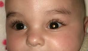
Leukokoria disebut juga pupil putih atau mata kucing. Leukokoria merupakan refleks pupil yang tidak normal, di mana refleks pupil berwarna putih. Leukokoria ini sering merupakan tanda pertama dari berbagai gangguan mata yang serius. Leukokoria umumnya terlihat pada katarak kongenital, penyakit Coats, retinoblastoma, retinopati prematuritas, infeksi toksokariasis, dan lain lain. Leukokoria merupakan tanda yang mengkhawatirkan terutama di kelompok usia anak-anak yang harus segera dideteksi dan diobati.
Penyebab
Leukokoria merupakan kelainan dari refleks pupil akibat adanya penyumbatan dari pembuluh darah sekitar retina yang direfleksikan melalui pupil. Beberapa penyakit yang dapat menyebabkan leukokoria, seperti infeksi pada bagian mata, contohnya uveitis, endoftalmitis, infeksi toksokariasis kekeruhan lensa seperti katarak, persistent hyperplastic primary vitreous (PHPV), kelainan retina seperti retinoblastoma, pengelupasan retina atau retinal detachment, retinopati prematuritas, miopia tinggi, koloboma, penyakit Coats, dan kelainan bagian dalam mata lainnya.
Faktor Risiko
Faktor risiko terjadinya leukokoria yaitu adanya faktor genetik keluarga, riwayat penyakit yang berhubungan dengan leukokoria di keluarga, adanya riwayat kelahiran prematur dapat mengindikasikan adanya retinopati prematuritas. Leukokoria akibat retinopati prematuritas muncul akibat adanya pengelupasan retina. Trauma saat lahir juga dapat menjadi salah satu faktor risiko terjadinya leukokoria karena adanya perdarahan di dalam bola mata.
Gejala
Leukokoris ditandai dengan adanya refleks pupil yang keputihan, pupil dapat terlihat normal pada cahaya kamar namun tidak memiliki red reflex pada pemeriksaan oftalmoskop. Gambaran leukokoria awalnya paling sering disadari oleh keluarga sebagai mata kucing atau melalui kilatan cahaya foto.
Diagnosis
Setiap orang dengan leukokoria harus dievaluasi secara anamnesis atau wawancara lebih dalam mengenai gejala lain, riwayat penyakit dahulu, riwayat lahir dan riwayat keluarga, selanjutnya perlu dilakukan pemeriksaan fisik lebih lanjut seperti refleks pupil, pemeriksaan dengan menggunakan slit lamp, oftalmoskopi, ultrasonografi, dan lain-lain.
Pemeriksaan refleks pupil dilakukan di ruangan gelap, dilihat menggunakan oftalmoskop. Ketika cahaya diproyeksikan ke mata, refleks merah normal harus berasal dari kedua mata dan harus simetris dalam warna dan kontrasnya. Refleks dianggap abnormal apabila refleks berkurang, refleks putih, terdapat bitnik gelap pada refleks atau asimetri (refleks Bruckner).
Jika hasil pemeriksaan refleks mata meragukan, dapat ditetesi obat untuk dilatasi pupil terlebih dahulu sebelum pemeriksaan. Warna dari refleks pupil juga dapat menjadi ciri khas dari suatu penyakit seperti pada retinoblastoma ditandai dengan refleks pupil keputihan, pada katarak kongenital ditandai dengan refleks abu kebiruan, pada penyakit Coats dan pengelupasan retina ditandai dengan refleks kuning.
Pemeriksaan selanjutnya dapat dilakukan ultrasonografi, pemeriksaan ini direkomendasikan pada semua pasien dengan leukokoria karena sangat spesifik untuk mendeteksi kalsifikasi intraokular (90%) pada retinoblastoma. Retinoblastoma terlihat sebagai massa padat di retina dengan kemampuan merefleksikannya yang tinggi karena kalsifikasi.
Pemeriksaan Optical Coherence Tomography (OCT) adalah pemeriksaan noninvasif untuk menilai cairan, pembengkakan, atau fibrosis pada bagian belakang mata. Pemeriksaan menggunakan CT-scan dihindari pada anak-anak karena paparan radiasi yang tinggi, tetapi dapat berguna jika terdapat kalsifikasi intraokular yang meragukan. Pemeriksaan dengan menggunakan MRI adalah modalitas diagnostic pilihan untuk mendeteksi penyebaran intrakranial retinoblastoma dan tumor sekunder lainnya seperti pinealoblastoma atau neuroblastoma parasellar.
Pemeriksaan darah dapat dilakukan guna melihat apakah terdapat infeksi TORCH (Toxoplasma gondii, Rubella, CMV, Herpes simplex virus). Kemudian ada juga pemeriksaan tes genetic yang membantu dalam mendiagnosis retinoblastoma, penyakit coats, dan penyakit genetik lainnya.
Tata Laksana
Leukokoria merupakan tanda dari penyakit mata, sehingga tata laksana leukokoria tergantung dari diagnosis penyakitnya. Apabila leukokoria disebabkan oleh retinoblastoma, maka dapat diterapi menggunakan beberapa pilihan metode, seperti:
- Enukleasi, yaitu pembedahan yang mengangkat keseluruhan bola mata dapat disertai atau tidak disertai dengan konjungtiva, dilakukan pada mata yang buta dan nyeri dengan melibatkan eksisi sebagian saraf optik untuk mencegah penyebaran intrakranial.
- Kemoreduksi, yaitu terapi untuk mengurangi ukuran tumor.
- Termoterapi, yaitu terapi dengan diberikan panas dari sinar infra merah sehingga memicu kematian sel tumor. Termoterapi menggunakan panas dengan suhu 40 C hingga 60 C.
- Krioterapi, yaitu terapi yang bekerja dengan cara merusak lapisan pembungkus sel tumor melalui cara dibekukan.
- Fotokoagulasi laser, yaitu terapi untuk retinoblastoma dengan prinsip kerjanya menghasilkan skar akibat energi panas yang bersumber dari sinar laser. Tumor akan dilapisi dengan skar berlapis untuk membatasi aliran darahnya. Indikasi fotokoagulasi laser ini adalah tumor besar yang telah dilakukan kemoreduksi.
- Radioterapi, yaitu salah satu modalitas penting dalam tatalaksana retinoblastoma. Radioterapi digunakan pada tumor yang telah mengecil oleh kemoreduksi atau muncul kembali setelah kemoterapi.
Selain retinoblastoma, leukokoria juga dapat disebabkan oleh penyakit Coats. Pada penyakit Coats, dapat ditata laksana dengan laser atau krioterapi untuk mengeluarkan cairan di retina. Apabila leukokoria disebabkan oleh katarak, dapat dilakukan tindakan pembedahan. Katarak yang dioperasi pada usia antara 2 bulan hingga 6 bulan memberikan hasil yang baik.
Pada kasus infeksi toksokariasis, obat-obatan jenis kortikosteroid adalah pengobatan utama dalam mengendalikan peradangan. Peran terapi anti cacing pada toksokariasis mata masih menjadi kontrovesi dan harus selalu menyertai steroid. Pada penyakit retinopati prematuritas, observasi, laser retina, krioterapi dan operasi retina adalah beberapa terapi pilihan untuk retinopati prematuritas. Pembuluh darah retina yang imatur dapat diamati dan di evaluasi berulang.
Komplikasi
Komplikasi yang dapat terjadi pada leukokoria bermacam-macam tergantung dari diagnosisnya. Pada kasus retinoblastoma, komplikasi yang mungkin terjadi seperti invasi tumor ke sklera dan koroid, metastasis ke tempat yang lebih jauh, dan kematian. Pada kasus uveitis atau infeksi mata pada anak, komplikasi yang memungkinkan terjadi seperti ambliopia (mata malas), katarak, glaukoma, dan cystoid macular edema. Katarak dan operasi katarak juga memiliki komplikasi seperti ambliopia, anisometria, capsular opacification, glaukoma sekunder, uveitis, endoftalmitis pasca operasi, dan pelepasan retina (retinal detachment). Komplikasi yang sering terjadi dari termoterapi yaitu atrofi iris atau iris mengecil, kekeruhan lensa paraksial, dan pelepasan retina.
Pencegahan
Kurangnya pengetahuan dan kewaspadaan mengenai leukokoria mengakibatkan pasien terlambat didiagnosis. Hal ini menjadi hambatan terbesar dalam meningkatkan angka kesembuhan pasien dengan leukokoria. Anak dengan riwayat keluarga retinoblastoma, riwayat kelahiran premature dan riwayat terinfeksi TORCH membutuhkan pengawasan lebih ketak serta evaluasi berkala untuk menemukan kelainan lebih awal sehingga dapat diterapi secepatnya.
Kapan Harus ke Dokter?
Apabila terdapat bintik putih pada bagian hitam pada mata anak, keluhan tidak bisa melihat, terdapat cahaya putih di mata saat difoto, seperti mata kucing, anak kesulitan mengerjakan aktivitas sehari-hari seperti menonton tv terlalu dekat, sering terjatuh atau terbentur, segera konsultasikan anak anda ke dokter mata.
Mau tahu informasi seputar penyakit lainnya? Cek di sini, ya!
- dr Ayu Munawaroh, MKK
Diagne JP, Sow AS, Ka AM, Wane AM, Ndoye Roth PA, Ba EA, De Medeiros ME, Ndiaye JM, Diallo HM, Kane H, Sow S, Nguer M, Sy EM, Ndiaye PA. [Rare causes of childhood leukocoria]. J Fr Ophtalmol. 2017 Oct;40(8):676-680.
A Stepwise Approach to Leukocoria [Internet]. American Academy of Ophthalmology. 2021
Leukocoria - American Association for Pediatric Ophthalmology and Strabismus [Internet]. Aapos.org. 2021