Definisi
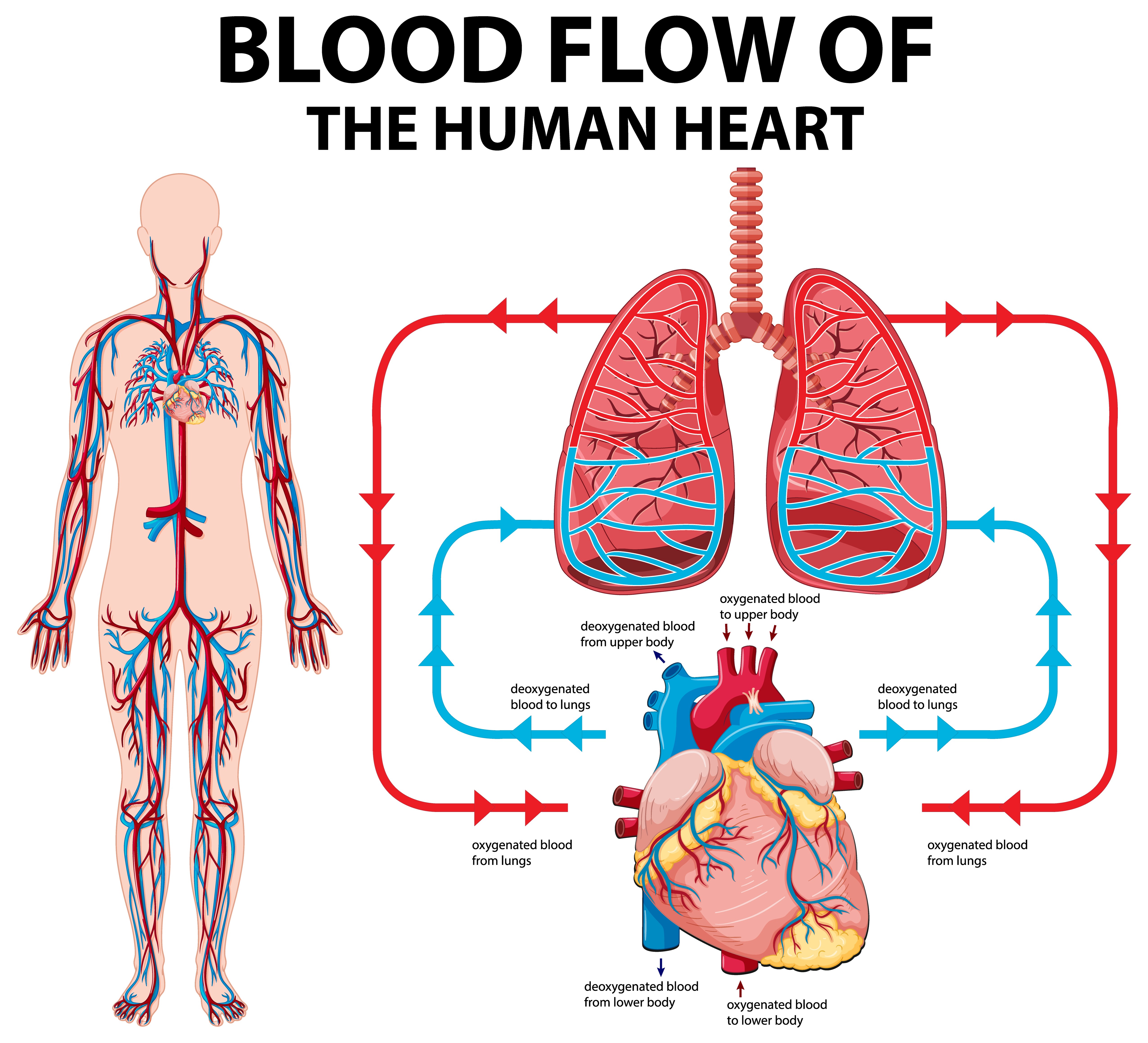
Oklusi Arteri retina disebut juga central retinal artery occlusion (CRAO) merupakan penyakit mata yang ditandai dengan tersumbatnya pembuluh darah arteri retina secara mendadak. Retina merupakan lapisan tipis pada bagian belakang mata yang memiliki peran untuk mengubah cahaya menjadi sinyal saraf agar dapat dibaca oleh otak. Di retina terdapat dua pembuluh darah utama yaitu arteri retina sentral dan vena retina sentral. Arteri pada retina membawa darah yang kaya oksigen untuk retina. Jika terjadi penyumbatan pada arteri utama atau pada cabang kecil, akan menyebabkan sel kekurangan oksigen sehingga retina akan berangsur-angsur mengalami kerusakan. Sumbatan mendadak yang terjadi pada arteri akan mengakibatkan hilangnya penglihatan secara mendadak. Oklusi arteri retina, merupakan kasus kegawatdaruratan dan keterlambatan penanganan akan mengakibatkan kebutaan yang permanen.
Penyebab
Penyebab oklusi arteri retina bermacam-macam meliputi hipertensi tidak terkontrol, diabetes melitus, penyakit katup jantung, penyakit jantung bawaan, pengguna narkoba jarum suntik, dan kondisi pengentalan darah. Selain itu, riwayat cedera, kelainan bawaan dari lahir pada arteri retina sentral, perlambatan aliran pembuluh darah akibat peningkatan tekanan intraokular, stenosis arteri juga dapat menyebabkan oklusi arteri retina. Plak yang menyumbat arteri retina dapat berasal dari kalsium , kolesterol, akibat operasi jantung, ataupun karena bakteri.
Faktor Risiko
Penyakit oklusi arteri retina lebih banyak dialami oleh laki-laki dibandingkan perempuan. Biasanya penyakit ini dialami oleh orang berusia diatas 60 tahun, namun pada beberapa kasus dapat dijumpai pada penderita yang lebih muda hingga usia 30 tahun. Oklusi arteri retina ini biasanya hanya mengenai satu mata saja, namun 1-2% penderita ditemukan gangguan pada kedua mata. Mata kanan dan kiri memiliki kesempatan terkena yang sama. Kasus oklusi arteri retina ini lebih sedikit dibandingkan dengan oklusi vena retina.
Gejala
Gejala utama dari oklusi arteri retina adalah :
- Ketajaman penglihatan turun secara mendadak.
- Penglihatan tiba-tiba gelap tanpa terlihatnya kelainan pada mata luar. Biasanya hal ini hanya terjadi pada satu sisi mata.
- Gangguan penglihatan tidak disertai dengan keluhan gatal atau nyeri.
- Penglihatan kabur terkadang bersifat hilang timbul.
- Reaksi pupil pada kasus oklusi arteri retina menjadi lemah dengan pupil tidak simetris.
- Gangguan penglihatan yang terjadi biasanya cukup berat.
Penderita oklusi arteri retina harus mewaspadai adanya sumbatan mendadak di organ lainnya seperti sumbatan mendadak di jantung yang menyebabkan serangan jantung, atau sumbatan di otak yang mengakibatkan stroke.
Diagnosis
Diagnosis oklusi arteri retina dapat ditegakkan melalui rangkaian wawancara mendalam (anamnesa), pemeriksaan visus dan funduskopi. Gejala umum pada oklusi arteri retina adalah hilangnya atau terganggunya penglihatan tanpa rasa nyeri. Pada oklusi arteri retina umumnya penurunan tajam penglihatan terjadi pada satu mata, berat dan dalam waktu cepat dan mendadak. Sebagian besar pasien melaporkan adanya riwayat episode hilang/gangguan fungsi penglihatan sebelumnya secara transien yaitu membaik kembali, dalam hitungan detik hingga menit dan membaik dengan sendirinya. Pada beberapa kasus, dapat didahului dengan gejala yang mengindikasikan adanya oklusi seperti nyeri kepala, nyeri pada leher, sensasi tidak nyaman di kulit kepala. Gejala-gejala ini dapat berlangsung bersamaan atau mendahului gejala gangguan penglihatan akibat oklusi. Riwayat menderita penyakit sistemik yang dapat membentuk emboli (sumbatan pembuluh darah) penting dalam menegakkan diagnosis. Penderita memerlukan pemeriksaan tekanan darah, elektrokardiografi, kadar gula darah, kadar lemak dan kolesterol untuk mendeteksi penyakit sistemik seperti hipertensi, aterosklerosis atau diabetes.
Pada pemeriksaan refleks pupil akan ditemukan adanya refleks pupil tidak normal pada mata yang sakit, selain itu pemeriksaan bagian depan mata akan tampak normal kecuali bila sudah terjadi komplikasi. Pada pemeriksaan funduskopi akan memberikan gambaran khas yang disebut cherry red spot. Namun pada pemeriksaan funduskopi pun dapat terlihat normal dalam menit-menit pertama sampai beberapa jam setelah oklusi terjadi. Pemeriksaan penunjang dibutuhkan seperti electroretinography, collor doppler adalah salah satu bentuk ultrasonografi dimana pada oklusi arteri retina dapat menunjukkan adanya penurunan atau hilangnya kecepatan aliran darah pada arteri retina sentral.
Tata Laksana
Hingga kini, pengobatan yang tepat untuk memulihkan penglihatan setelah terjadi oklusi arteri retina masih dalam tahap penelitian. Penanganan terhadap penderita oklusi arteri retina direkomendasikan dalam waktu 24 jam setelah munculnya penurunan tajam penglihatan. Ada beberapa pilihan pengobatan yang dapat dilakukan seperti masase bola mata, yaitu menekan bola mata selama 5-15 detik lalu tekanan dilepaskan. Hal ini dapat dilakukan beberapa kali dengan tujuan melepaskan sumbatan arteri yang terjadi. Tindakan pemijatan ini akan membuat arteri retina melebar yang secara teori meningkatkan perfusi (aliran darah) retina. Parasentesis yaitu tindakan mengeluarkan sebagaian cairan di dalam bola mata. Teknik parasentesis ini akan menyebabkan penurunan tekanan bola mata secara tiba-tiba dengan tujuan agar tekanan perfusi arteri di belakang sumbatan dapat mendorong emboli ke perifer (tepi). Obat-obatan dapat diberikan dengan tujuan menurunkan tekanan bola mata seperti tetes mata Timolol dan obat sistemik yaitu acetazolamide. Laser Nd:YAG dapat dilakukan dengan tujuan membantu menghancurkan sumbatan arteri retina. Namun laser ini juga memiliki komplikasi seperti terbentuknya aneurisma (pertumbuhan tidak normal pembuluh darah) dan perdarahan vitreus. Terapi hiperbarik dilakukan dengan memberikan oksigen 100% dalam tekanan tinggi. Terapi ini efektif pada oklusi arteri retina yang terjadi kurang dari 12 jam.
Komplikasi
Komplikasi oklusi arteri retina adalah neovaskularisasi iris dan neovaskularisasi diskus optik. Neovaskularisasi merupakan pertumbuhan pembuluh darah baru dimata sebagai akibat dari keadaan kurang oksigen. Penderita oklusi arteri retina dianjurkan kontrol ulang secara rutin selama 3 bulan pertama sehubungan dengan risiko komplikasi. Selain komplikasi pada mata, perlu diwaspadai juga bahwa pasien dengan kelainan oklusi pembuluh darah retina mempunyai risiko 10% terkena stroke pada tahun pertama penyakit dan akan terus meningkat.
Pencegahan
Beberapa langkah dapat dilakukan untuk mencegah terjadinya oklusi arteri retina seperti menjaga tekanan darah terkontrol dibawah 140/90 mmHg, menjaga kolesterol LDL tetap normal, mengonsumsi makanan tinggi serat dan rendah lemak, berolahraga dengan teratur. Jika Anda menderita diabetes, maka Anda harus rutin minum obat dan melakukan kontrol secara rutin ke dokter agar gula darah tetap terpantau.
Kapan harus ke dokter?
Disarankan untuk segera ke dokter apabila mengalami penurunan ketajaman penglihatan mata mendadak tanpa disertai rasa nyeri. Penurunan ketajaman penglihatan yang mendadak pada beberapa kasus dapat hilang timbul sehingga disarankan untuk segera ke dokter agar mendapaatkan penanganan dini. Disarankan untuk rutin melakukan pemeriksaan mata apabila memiliki faktor risiko seperti tekanan darah tinggi, riwayat kolesterol tidak terkontrol, diabetes melitus, riwayat pengentalan darah, dan riwayat cedera.
- dr Nadia Opmalina
American Academy of Ophthalmology. Retinal and Ophthalmic Artery Occlusions Preferred Practice Pattern. AAO: Elsevier Inc. 2016. https://doi.org/10.1016/j.ophtha.2016.09.024
American Society of Retina Specialists. Retinal Artery Occlusion. 2017. https://www.asrs.org/content/documents/fact-sheet-29-retinal-artery-occlusion.pdf
American Academy of Ophthalmology. Diagnosis and Management of Central Retinal Artery Occlusion. AAO: Eyenet Magazine. 2017. https://www.aao.org/eyenet/article/diagnosis-and-management-of-crao
Medscape. Central Retinal Artery Occlusion [Updated 2019 Jun 11]. https://emedicine.medscape.com/article/1223625-overview#a6


