Definisi
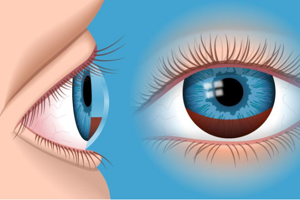
Perdarahan vitreus adalah perdarahan yang terjadi di dalam bola mata. Perdarahan ini biasanya tidak disertai dengan rasa nyeri, namun menyebabkan kehilangan penglihatan yang cukup signifikan. Angka kejadian perdarahan vitreus sangat tergantung dari penyebabnya, meskipun dapat terjadi secara spontan. Secara umum, angka kejadian perdarahan vitreus adalah sekitar 7 kejadian per 100.000 penduduk.
Penyebab
Vitreus merupakan cairan mirip gel yang mengisi bola mata, terdiri atas 99% air dan sisanya kolagen dan asam hialuronat. Vitreus berfungsi untuk mempertahankan bentuk bola mata, menangkap getaran saat ada hentakan pada mata, serta menjaga agar lapisan retina atau saraf pada mata tetap terhubung dengan bagian belakang mata. Vitreus berwarna bening sehingga dapat dilewati oleh cahaya yang masuk ke dalam bola mata. Di dekat vitreus, terdapat beberapa pembuluh darah yang mengalirkan darah untuk daerah retina. Pada perdarahan vitreus, pembuluh darah dapat pecah akibat benturan yang sangat keras, atau karena adanya struktur yang bermasalah.
Perdarahan vitreus dapat terjadi sebagai akibat dari kondisi tertentu atau dapat terjadi secara spontan. Penyebab tersering perdarahan vitreus adalah retinopati diabetik proliferatif, yaitu sebuah kondisi ketika retina mata mengalami kerusakan yang parah disebabkan oleh diabetes. Pembuluh darah di sekitarnya pun juga mengalami kerusakan, sehingga tubuh berusaha membentuk pembuluh darah baru agar retina tetap memiliki nutrisi dan oksigen yang cukup. Namun, pembuluh darah baru ini dapat tumbuh ke arah vitreus dan jauh lebih rapuh sehingga mudah pecah. Selain itu, penyebab lainnya dapat berupa robekan pada lapisan retina, serta adanya cedera pada bola mata.
Penyebab lainnya dapat berupa pecahnya aneurisma (atau pelebaran pembuluh darah disertai dengan penipisan lapisan pembuluh darah) dan adanya pembuluh darah baru yang disebabkan berbagai kondisi seperti degenerasi makula (penurunan fungsi saraf mata).
Faktor Risiko
Faktor risiko perdarahan vitreus adalah adanya penyakit diabetes yang tidak terkontrol dengan obat. Pada usia di bawah 40 tahun, perdarahan vitreus lebih sering terjadi akibat cedera pada bola mata, sementara di atas 40 tahun tanpa diabetes, dapat terjadi akibat adanya robekan pada retina. Selain itu, adanya kelainan pada darah seperti leukemia (kanker darah) dan trombositopenia (kadar trombosit yang terlalu rendah pada darah) dapat mempercepat terjadinya perdarahan vitreus. Perdarahan vitreus yang spontan dapat terkait dengan kerusakan pada pembuluh darah mata akibat penyakit jantung atau diabetes disertai penggunaan obat pengencer darah, atau akibat batuk yang terlalu keras serta angkat beban.
Gejala
Gejala yang umum dikeluhkan pada perdarahan vitreus adalah adanya kehilangan penglihatan pada satu mata secara tiba-tiba. Gejala ini dapat disertai dengan keluhan adanya persepsi “benda melayang”, penglihatan seperti diselimuti kabut, serta penglihatan seperti terhalang jaring laba-laba. Keluhan ini pada umumnya tidak disertai dengan nyeri.
Diagnosis
Perdarahan vitreus dapat diperiksa secara langsung menggunakan alat bernama funduskopi. Dengan alat tersebut, akan tampak darah melayang di ruang vitreus, menghalangi tampilan retina secara keseluruhan. Sebagai dampak dari perdarahan vitreus, fungsi mata seperti ketajaman penglihatan dan refleks cahaya dapat pula diperiksa. Ketajaman penglihatan biasanya mengalami penurunan akibat darah yang menghalangi cahaya masuk ke dalam bola mata. Pada kasus yang dicurigai akibat cedera, dokter dapat melakukan pemeriksaan slit lamp untuk mencari adanya perdarahan pada bola mata bagian depan. Dokter juga dapat melakukan pemeriksaan tekanan darah karena tekanan darah yang terlalu tinggi juga dapat menyebabkan pembuluh darah pada mata pecah.
Pemeriksaan laboratorium dan pencitraan pada umumnya dilakukan untuk mengetahui penyebab perdarahan vitreus. Pemeriksaan laboratorium yang dapat dilakukan adalah pemeriksaan gula darah, darah perifer lengkap, waktu pembekuan darah, serta apus darah tepi. Sementara itu, pencitraan yang dapat dilakukan dapat berupa USG mata untuk melihat karakteristik perdarahan, optical coherence tomography (OCT) untuk membedakan asal pembuluh darah yang pecah, serta CT scan atau magnetic resonance imaging (MRI) untuk mencari perdarahan otak yang dapat memengaruhi perdarahan vitreus.
Tata Laksana
Tata laksana perdarahan vitreus bertujuan untuk menangani perdarahan serta mengatasi penyebab. Vitrektomi, atau pembedahan untuk mengeluarkan cairan vitreus, dapat dilakukan jika retina tidak dapat terlihat dengan baik. Jika terdapat pembuluh darah baru pada bagian depan bola mata, dokter dapat menyarankan Anda untuk menjalani pembedahan terkait.
Selain itu, terapi yang bertujuan untuk menghentikan perdarahan dari pembuluh darah dapat berupa fotokoagulasi laser, yaitu teknik pembedahan menggunakan laser untuk menutup pembuluh darah yang pecah atau bocor. Jika dalam waktu 1 bulan vitreus belum kembali jernih, dokter akan melakukan vitrektomi.
Selain terapi di atas, terapi menggunakan obat dapat bertujuan untuk menurunkan pertumbuhan pembuluh darah baru pada mata yang berpotensi rapuh dan mudah pecah.
Hal yang perlu diperhatikan saat mengalami perdarahan vitreus adalah sebagai berikut:
- Menurunkan aktivitas fisik yang berat, seperti angkat beban. Hal ini menyebabkan kenaikan tekanan darah secara tiba-tiba, yang dapat memicu perdarahan vitreus kembali.
- Tidur dengan posisi kepala terangkat 45 derajat, agar darah menetap di bagian bawah bola mata sehingga cahaya dapat masuk ke dalam bola mata dengan baik.
- Jika Anda mengonsumsi obat pengencer darah, konsultasi terlebih dahulu kepada dokter Anda apakah konsumsi perlu dihentikan. Jika tidak ada kerusakan signifikan pada mata sementara obat pengencer darah perlu diberikan, biasanya dokter akan meminta Anda untuk tetap mengonsumsi pengencer darah.
Komplikasi
Komplikasi perdarahan vitreus dapat berupa hemosideris bulbi, atau kerusakan struktur mata karena terikat dengan ion besi. Pada perdarahan vitreus, sel darah merah dapat dirombak kembali, salah satunya menjadi ion besi. Ion besi ini kemudian berikatan permanen dengan struktur pada bola mata, sehingga menyebabkan kerusakan pada struktur tersebut.
Selain itu, komplikasi lainnya adalah glaukoma. Glaukoma dapat terjadi karena komponen sel darah merah bernama hemoglobin dapat menghalangi jalur pengeluaran cairan di bagian depan bola mata, sehingga aliran cairan tidak lancar dan menyebabkan tekanan bola mata meningkat. Selain itu, komponen lainnya berupa ion besi dapat berikatan pada saluran tersebut sehingga terjadi kerusakan permanen.
Jika perdarahan vitreus terjadi pada bayi, komplikasi lanjutan dapat berupa ambliopia (mata malas) dan miopia (rabun jauh). Oleh karena itu, perdarahan vitreus pada bayi harus segera dioperasi.
Pencegahan
Pencegahan perdarahan vitreus dapat berupa kontrol gula darah dan tekanan darah dengan pola makan sehat, aktivitas fisik teratur, serta menjaga berat badan ideal. Selain itu, jika Anda telah diketahui mengalami diabetes atau hipertensi, sebaiknya Anda patuh dalam mengonsumsi obat agar kadar gula darah atau tekanan darah terkontrol. Sebagai tambahan, beberapa penelitian menunjukkan adanya keuntungan dalam pemberian obat pencegah pembuluh darah tumbuh pada pasien yang baru saja dioperasi akibat perdarahan vitreus, agar perdarahan vitreus tidak terjadi kembali.
Kapan Harus ke Dokter?
Jika Anda merasa penglihatan Anda tiba-tiba buram tanpa disertai rasa nyeri atau mata merah, segeralah ke dokter. Penyebab mata buram mendadak bukan hanya perdarahan vitreus, namun banyak di antaranya yang terkait dengan kerusakan retina atau pembuluh darah, sehingga membutuhkan penanganan segera.
Mau tahu informasi seputar penyakit lainnya? Cek di sini, ya!
- dr Nadia Opmalina
Jena, S., & Tripathy, K. (2021). Vitreous Hemorrhage. Retrieved 22 November 2021, from https://www.ncbi.nlm.nih.gov/books/NBK559131/
Johnson, B., Feldman, B., Shah, V., Reed, D., Tripathy, K., & Weng, C. (2021). Vitreous Hemorrhage - EyeWiki. Retrieved 22 November 2021, from https://eyewiki.aao.org/Vitreous_Hemorrhage
Phillpotts, B. (2018). Vitreous Hemorrhage: Background, Pathophysiology, Epidemiology. Retrieved 22 November 2021, from https://emedicine.medscape.com/article/1230216-overview