Definisi
Retinitis pigmentosa (RP) adalah kumpulan kelainan genetik yang melibatkan kerusakan dan kematian sel yang menerima cahaya di retina. Sel ini bersifat sensitif terhadap cahaya dan berada di bagian belakang mata. RP adalah penyakit langka yang menyerang satu dari 4.000 orang di Amerika Serikat dan di seluruh dunia. Data penelitian di Asia Selatan menunjukkan bahwa terdapat lebih dari setengah gen pada penduduk Asia Selatan khususnya daerah Pakistan dan India yang memiliki keterkaitan dengan RP.
Penyebab
Retinitis Pigmentosa (RP) merupakan penyakit yang diwariskan secara genetik dan menyebabkan sel yang menerima cahaya atau disebut fotoreseptor rusak atau tidak dapat berfungsi dengan baik. Sebagian besar kasus RP disebabkan oleh mutasi pada gen rhodopsin. Umur saat mulai terjadinya gejala RP, kecepatan dari perkembangan gejala, dan kehilangan fungsi penglihatan bergantung dari pola penurunan gen dalam keluarga masing-masing. Pada RP terjadi kerusakan pada sel fotoreseptor mengenai sel batang dan sel kerucut yang ada di retina. Sel batang berada pada lapisan luar dari retina, sel ini berfungsi untuk melihat dalam suasana remang dan gelap. Sel kerucut berfungsi untuk melihat dengan detail yang lebih jelas pada keadaan terang. Sebanyak 90 gen telah diidentifikasi dapat menyebabkan RP.
Faktor Risiko
Faktor risiko utama anda bisa mengalami RP adalah riwayat penyakit RP di keluarga. Pola genetik yang diturunkan pada setiap keluarga dapat berbeda bergantung dari jenis mutasi dan gen yang bermutasi.
Gejala
Pada tahap awal dari RP, sel batang akan mengalami kerusakan terlebih dahulu sehingga gejala yang dirasakan adalah lapang pandang mata yang menyempit hingga lapang pandang mata seperti melihat menggunakan terowongan (tunnel vision). Gejala lain yang dapat anda alami seperti miopia atau rabun jauh. Pada beberapa pasien, tajam penglihatan bisa normal. Kecepatan perkembangan gejala berbeda pada setiap individu. Umur awal terjadinya gejala awal RP berbeda pada setiap orang. Sebagian besar individu mengalami kebutaan pada umur 40 tahun dari perjalanan penyakit RP. Perubahan anatomis atau bentuk dari retina dan perkembangan kerusakan anatomis yang dialami harus dinilai menggunakan oftalmoskop oleh dokter spesialis mata.
Diagnosis
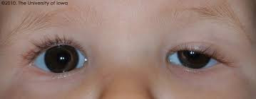
Diagnosis RP ditegakkan oleh dokter menggunakan oftalmoskopi. Ofalmoskopi adalah alat untuk melihat bagian fundus atau bagian belakang mata. Bagian mata yang dapat dilihat menggunakan oftalmoskop termasuk retina, koroid, pusat saraf mata, dan pembuluh darah. Pada pemeriksaan oftalmoskopi akan terlihat gambaran bintik kegelapan yang merupakan deposit pigmen di retina. Pemeriksaan lain yang dapat dilakukan oleh dokter untuk menegakkan diagnosis RP adalah:
- Elektroretinogram (ERG): Fungsi alat ini adalah untuk mengukur aktifitas listrik yang ada pada sel di retina. Pemeriksaan ini sensitif dalam memeriksa RP.
- Pemeriksaan lapang pandang: Pemeriksaan ini digunakan untuk menilai derajat keparahan atau besar kerusakan sel fotoreseptor di retina terhadap lapang pandang anda. Pemeriksaan dapat dilakukan dengan dua cara yaitu dengan tes konfrontasi dan pemeriksaan menggunakan alat perimetri. Tes konfrontasi menggunakan acuan lapang pandang dokter yang dibandingkan dengan lapang pandang pasien. Pemeriksaan perimetri menggunakan cahaya berwarna merah yang akan ditembakkan dan pasien akan menekan tombol jika melihat cahaya tersebut.
- Pemeriksaan genetik: dilakukan pada beberapa orang untuk memeriksa mutasi genetik. Informasi mengenai mutasi genetik penting untuk mengetahui pola keturunan dari RP dalam keluarga.
- Pemeriksaan untuk mendeteksi penyakit lain yang menyerupai RP seperti sifilis. Pemeriksaan sifilis menggunakan pemeriksaan darah pasien.
Tata Laksana
Tatalaksana untuk RP meliputi deteksi dini dan pengawasan perkembangan gejala secara teratur. Hingga saat ini, masih belum ada terapi yang pasti untuk mengobati dan menghentikan perjalanan penyakit RP.
- Pemeriksaan tahunan disarankan untuk mendeteksi komplikasi yang dapat menganggu penglihatan dan mengetahui perkembangan penyakit.
- Jika anda mengalami komplikasi dari RP seperti katarak, operasi katarak dapat membantu fungsi penglihatan mata.
- Hindari merokok, merokok dapat mempercepat perkembangan penyakit RP dibandingkan dengan mereka yang tidak merokok.
- Penggunaan kacamata yang dapat menghalangi sinar matahari hingga 550 nanometer yang disertai dengan kaca pelindung di bagian samping disarankan.
- Penggunaan suplementasi vitamin A atau penggunaan obat lain dikonsumsi sesuai dengan anjuran dokter.
- Beberapa obat seperti asetazolamide oral dan inhibitor karbonik anhidrase topikal dapat membantu mengurangi gejala RP.
- Obat-obatan lain seperti antagonis kalsium menunjukkan beberapa keuntungan namun efikasi dan keamanan dari obat ini belum sepenuhnya dimengerti.
- Pengobatan yang bersifat retinatoksik harus dihentikan dan digunakan dengan hati-hati pada pasien dengan RP. Beberapa obat termasuk obat untuk megnobati disfungsi ereksi, isoretinoin dan retinoid lainnya. Penggunaan obat-obatan ini dan obat yang bersifat neurotoksik harus digunakan sesuai dengan anjuran dan pengawasan dokter.
Komplikasi
Beberapa komplikasi yang dapat dtiemukan pada pasien dengan RP adalah katarak, glaukoma, keratokonus, dan lepasnya bagian vitreous yang menempel pada bagian belakang retina (posterior vitreous detachment).
- Katarak: Katarak dalah keadaan dimana lensa mata menjadi lebih opak secara progresif yang mmenyebabkan penglihatan yang kabur. Katarak pada pasien dengan RP disebabkan oleh proses inflamasi yang disebabkan oleh RP. Jenis katarak yang paling umum terjadi pada pasien dengan RP adalah katarak subkapsular posterior.
- Glaukoma: Glaukoma adalah gangguan yang terjadi pada beberapa bagian mata dan disebabkan oleh kenaikan tekanan bola mata.glaukoma pada kasus RP dikaitkan dengan migrasi atau perpindahan dari pigmen ke bilik mata bagian belakang dan menghalangi perpindahan cairan di mata.
- Posterior Vitreous Detachment (PVD): Terjadinya akumulasi cairan disebabkan oleh lapisan retina yang lepas dan penumpukkan cairan di bagian belakang retina. Akumulasi cairan akan merobek retina dari lapisan mata dan menyebabkan hilangnya fungsi penglihatan mata.
Pencegahan
Hingga saat ini belum ditemukan cara untuk mencegah perkembangan atau timbulnya RP. Hal ini dikarenakan RP lebih disebabkan oleh kelainan genetik yang teradi pada retina. Oleh karena itu, pemeriksaan secara berkala cukup penting dalam mengetahui gejala dini retinopati pigmentosa.
Kapan Harus ke Dokter?
Segera ke dokter jika memiliki gejala sebagai berikut:
- Penglihatan yang memburuk pada saat malam hari atau di lingkungan yang tidak memiliki penerangan yang baik.
- Penglihatan yang terganggu saat melihat benda-benda di lingkungan yang terang.
- Hilangnya penglihatan di sisi samping atau melihat seperti dari lubang kunci.
- Kesulitan dalam menilai kedalaman permukaan benda seperti saat sedang berjalan di tepi jalan atau menaiki tangga.
Mau tahu informasi seputar penyakit lainnya? Cek di sini, ya!
- dr Ayu Munawaroh, MKK
What Is Retinitis Pigmentosa?. American Academy of Ophthalmology. (2021).
Zafar, S., Ahmed, K., Ali, A., & Baig, R. (2017). Retinitis pigmentosa genes implicated in South Asian populations: a systematic review. Journal of the Pakistan Medical Association, 67(11), 1734.
Retinitis Pigmentosa | UVA Health [Internet]. Uvahealth.com. 2021
Encyclopedia M. Ophthalmoscopy: MedlinePlus Medical Encyclopedia [Internet]. Medlineplus.gov. 2021
Encyclopedia, M., & pigmentosa, R. (2021). Retinitis pigmentosa: MedlinePlus Medical Encyclopedia. Medlineplus.gov.
Kanski, J. J. (2020). Clinical diagnosis in ophthalmology. Philadelphia: Elsevier.
Smoking. Retina UK. (2021).
Glaucoma - Symptoms and causes. Mayo Clinic. (2021).
Rhegmatogenous Retinal Detachment (RRD): Background, Pathophysiology, Epidemiology. Emedicine.medscape.com. (2021).