Definisi
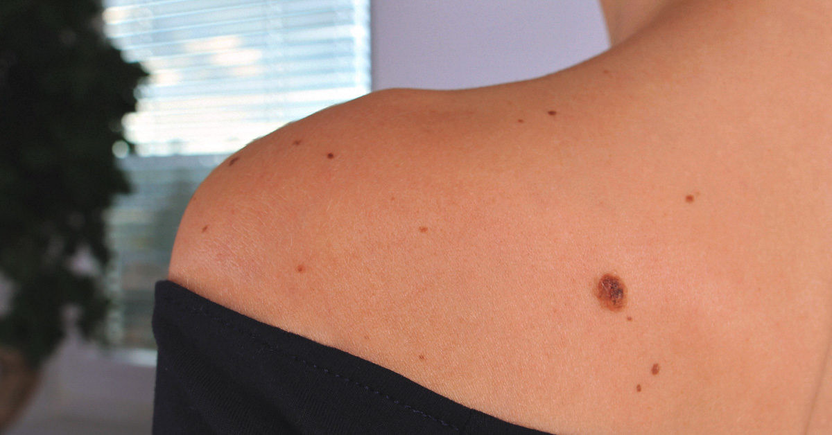
Nevus pigmentosus (jamak: nevi) adalah istilah medis untuk tahi lalat. Nevus ini terdiri dari kumpulan sel melanosit (sel pigmen kulit) yang jinak dan merupakan kondisi kulit yang sangat sering ditemukan. Nevus pigmentosus merupakan salah satu dari beberapa tipe nevus. Nama lain nevus pigmentosus adalah nevus melanositik, nevus nevositik.
Nevus pigmentosus dapat timbul sejak lahir (nevus pigmentosus kongenital) atau saat usia tertentu (nevus didapat). Terdapat beberapa jenis nevus kongenital dan nevus didapat.
Penyebab
Penyebab pasti pertumbuhan lokal dari sel melanosit sehingga membentuk nevus masih belum diketahui. Namun, yang pasti adalah jumlah nevus pigmentosus yang dimiliki seseorang bergantung pada faktor genetik, paparan sinar matahari, dan status imun. Hal ini dapat dijelaskan dengan:
- Orang yang memiliki banyak nevus pigmentosus biasanya memiliki anggota keluarga yang juga memiliki kondisi yang sama
- Pada nevus pigmentosus kongenital, telah terbukti bahwa ada mutasi atau kelainan pada gen tertentu.
- Pemakaian obat-obatan seperti vemurafenib, dabrafenib, serta obat penekan sistem imun dapat memicu timbulnya nevus pigmentosis baru.
Faktor Risiko
Hampir semua orang memiliki minimal satu nevus pigmentosus. Sekitar 1% populasi terlahir dengan satu atau lebih nevus pigmentosus kongenital yang kebanyakan tidak diturunkan. Sementara itu, orang kulit putih cenderung untuk memiliki lebih banyak nevus pigmentosus dibandingkan dengan orang berkulit gelap.
Nevus pigmentosus yang timbul saat masa kanak-kanak (usia 2 sampai 10 tahun) cenderung paling tampak dan menetap sepanjang umur. Sementara itu, nevus pigmentosus yang timbul pada masa kanak-kanak akhir atau pada usia dewasa biasanya akibat paparan sinar matahari dan dapat memudar dan mengecil seiring berjalannya waktu.
Gejala
Bentuk nevus pigmentosus dapat bervariasi. Berikut ini adalah ciri-ciri dari nevus pigmentosus:
- Nevus pigmentosus dapat timbul di bagian tubuh manapun
- Nevus dapat memiliki tampak yang berbeda pada bagian tubuh yang berbeda
- Nevus dapat datar atau menimbul
- Warna nevus dapat bervariasi dari pink, warna daging sampai cokelat tua, biru, atau hitam.
- Pada orang kulit putih, nevus cenderung untuk berwarna lebih terang. Sebaliknya, pada orang berkulit gelap, nevus cenderung untuk berwarna cokelat gelap sampai hitam
- Meskipun kebanyakan nevus pigmentosus berbentuk bulat atau oval, namun terkadang bentuknya bisa tidak seperti biasanya
- Ukuran nevus bervariasi dari. Diameter bisa dimulai dari beberapa milimeter sampai beberapa sentimeter
Diagnosis
Nevus pigmentosus biasanya didiagnosa dari tampakannya. Jika terdapat keraguan dalam mendiagnosa, biasanya akan dilakukan pemeriksaan lain misalnya menggunakan dermatoskopi. Pemeriksaan ini terutama diperlukan jika:
- Nevus mengalami perubahan ukuran, bentuk, struktur, atau warna
- Adanya nevus baru yang timbul pada usia dewasa (>40 tahun)
- Nevus yang memiliki kharakteristik yang berbeda dengan nevus kebanyakan orang
- Memiliki karakteristik ABCDE (asymmetry, border irregularity, colour variation, diameter > 6 mm)
- Nevus yang berdarah, berkoreng, atau gatal
Jika terdapat perubahan di atas, dokter biasanya juga akan melakukan pemeriksaan biopsi kulit. Pemeriksaan ini adalah satu-satunya cara untuk mengkonfirmasi atau menyingkirkan adanya kanker kulit. Teknik yang digunakan adalah biopsi eksisi, yang dilakukan untuk sekaligus mengangkat sel kanker dan jaringan kulit di sekitarnya untuk memastikan tidak ada sel kanker yang tertinggal.
Tata Laksana
Kebanyakan nevus pigmentosus tidak berbahaya dan tidak membutuhkan pengobatan atau terapi. Namun, nevus dapat diangkat pada keadaan seperti:
- Nevus yang terlihat mencurigakan seperti bentuk kanker kulit
- Nevus yang menimbulkan ketidaknyamanan, misalnya yang mudah teriritasi oleh pakaian, sisir, atau alat cukur
- Jika Anda merasa nevus pigmentosis mengganggu penampilan Anda.
Ada beberapa cara untuk membuang nevus pigmentosus, yaitu:
- Shave biopsy. Dokter akan menggunakan alat cukur untuk mencukur lapisan atas dari kulit
- Punch biopsy. Dokter akan menggunakan punch tool spesial untuk mendapatkan sampel kulit yang mengandung lapisan atas dan dalam dari kulit
- Biopsi eksisi
- Destruksi electrosurgical
- Laser untuk mengurangi pigmen atau membuang rambut kasar di atas nevus
Komplikasi
Banyak orang yang khawatir akibat adanya tahi lalat karena pernah mendengar mengenai melanoma, pertumbuhan sel melanosit yang bersifat ganas dan merupakan penyebab paling sering dari kematian akibat kanker kulit.
Pada fase awal, melanoma dapat terlihat seperti nevus pigmentosus yang tidak berbahaya, namun seiring berjalannya waktu, strukturnya akan menjadi tidak beraturan dan cenderung untuk bertambah besar. Orang degnan jumlah nevus pigmentosus yang lebih banyak, terutama jika jumlahnya lebih dari 100 nevi, memang lebih berisiko untuk menjadi melanoma, dibandingkan dengan yang hanya memiliki sedikit nevi.
Namun, nevus pigmentosus terkadang mengalami perubahan akibat hal lain seperti setelah paparan sinar matahari atau selama kehamilan. Saat itu, nevus dapat membesar, mengecil, atau bahkan menghilang.
Pencegahan
Jumlah nevus pigmentosus dapat dibatasi dengan melindungi kulit dari paparan sinar matahari, yang dimulai sejak lahir. Tabir surya saja tidak cukup untuk mencegah timbunya nevus baru.
Pada semua usia, proteksi dari sinar matahari penting untuk mengurangi penuaan kulit dan risiko kanker kulit. Cara untuk proteksi kulit adalah:
- Menutup kulit. Gunakan topi, lengan panjang, dan rok atau celana panjang. Pilihlah bahan yang didesain untuk melindungi kulit dari matahari (UPF 40+) saat berada di luar ruangan.
- Pakai tabir surya pada area tubuh yang tidak dapat tertutup oleh pakaian. Pilihlah tabir surya spektrum luas dengan proteksi tinggi (SPF 50+). Aplikasikan tabir surya secara berkala pada area tubuh yang terpapar sinar matahari
Kapan Harus ke Dokter?
Jika Anda tidak yakin mengenai bercak atau perubahan warna yang ada pada kulit Anda, maka sebaiknya Anda berkonsultasi dengan dokter. Sementara itu, jika nevus Anda terlihat berubah, misalnya mengalami perubahan warna, bentuk, atau ukuran, sebaiknya Anda juga berkonsultasi ke dokter. Hal ini untuk memeriksa adanya kemungkinan keganasan kulit yang dapat diniai dengan pemeriksaan biopsi kulit.
Kanker kulit paling mudah diterapi jika terdiagnosa dengan cepat. Oleh karena itu, penting untuk mengetahui ciri-ciri kanker kulit. Dokter mengembangkan sistem yang disebut dengan metode ABCDE untuk membantu orang mengidentifikasi tanda-tanda kanker kulit. ABCDE tersebut terdiri dari:
- A: Asimetris. Amati apakah nevus berbentuk tidak simetris
- B:Border (batas). Nevus pigmentosus harusnya memiliki batas yang tegas dan jelas serta reguler.
- C:Color(warna). Amati apakah nevus memiliki warna yang tidak rata (mengandung beberapa warna). Selain itu, amati juga adanya perubahan warna nevus
- D: Diameter. Hati-hati jika ukuran nevus lebih besar daripada penghapus yang ada di pensil
- E:Evolving (berevolusi). Cari adanya perubahan ukuran, warna, bentuk, atau permukaan nevus. Amati juga apakah ada gejala seperti gatal atau berdarah pada nevus.
Periksalah sendiri kulit Anda setidaknya setiap satu bulan. Anda juga harus mengingat bahwa kanker kulit dapat timbul di area tubuh yang tidak terlihat. Oleh karena itu, gunakanlah cermin untuk membantu Anda memeriksa bagian tubuh yang kurang terjangkau.
Mau tahu informasi seputar penyakit lainnya? Cek di sini, ya!
- dr Anita Larasati Priyono
Nevus: Definition, Common Types, Photos, Diagnosis, and Treatment. Healthline. (2022). Retrieved 7 May 2022, from https://www.healthline.com/health/nevus#photos.
Moles (melanocytic naevi, pigmented nevi) | DermNet NZ. Dermnetnz.org. (2022). Retrieved 7 May 2022, from https://dermnetnz.org/topics/melanocytic-naevus.
Moles: Who gets and types. Aad.org. (2022). Retrieved 7 May 2022, from https://www.aad.org/public/diseases/a-z/moles-types.
Bodman, M., & Aboud, A. (2022). Melanocytic Nevi. Ncbi.nlm.nih.gov. Retrieved 7 May 2022, from https://www.ncbi.nlm.nih.gov/books/NBK470451/.


