Definisi
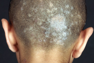
Tinea kapitis merupakan infeksi jamur pada kepala. Infeksi ini merupakan penyakit menular, dan terkait dengan infeksi jamur lainnya seperti jamur pada kaki (tinea pedis). Tinea kapitis juga merupakan infeksi yang cukup sering terjadi di seluruh dunia, terutama pada daerah yang panas dan lembab seperti Afrika, Asia Tenggara, dan Amerika Tengah.
Penyebab
Tinea kapitis disebabkan oleh kelompok jamur yang disebut sebagai dermatofita. Dermatofita membawahi tiga keluarga jamur, yaitu Tricophyton, Epidermophyton, dan Microsporum. Jamur-jamur ini menginfeksi bagian terluar kulit kepala dan rambut untuk memakan keratin yang ada pada kulit dan rambut. Hal ini dapat menyebabkan kerontokan atau kepatahan rambut. Penyebaran infeksi jamur dapat terjadi sebagai berikut:
- Orang ke orang (antropofilik). Tinea kapitis dapat ditularkan lewat kontak kulit dengan orang yang terinfeksi
- Hewan ke orang (zoofilik). Tinea kapitis juga dapat ditularkan dari hewan yang terinfeksi jamur tersebut. Jamur ini dapat menular ketika menyentuh atau memberikan perawatan pada kucing atau anjing yang terinfeksi. Jamur penyebab tinea kapitis sering ditemukan pada anak kucing, anak anjing, sapi, kambing, babi, dan kuda
- Tanah ke orang (geofilik). Jamur penyebab tinea kapitis juga dapat menyebar saat berkebun atau berkontak dengan tanah.
- Benda ke orang. Jamur penyebab tinea kapitis dapat menempel pada benda atau permukaan yang disentuh oleh orang atau hewan yang terinfeksi, seperti pakaian, handuk, seprai, sisir, dan sikat
Faktor Risiko
Faktor risiko tinea kapitis adalah:
- Usia. Tinea kapitis paling sering terjadi pada anak balita dan anak usia sekolah
- Paparan dengan anak lain. Wabah tinea kapitis sering terjadi di sekolah-sekolah serta tempat penitipan anak karena anak dapat saling berkontak saat bermain bersama
- Paparan terhadap hewan peliharaan. Hewan peliharaan seperti kucing dan anjing dapat terinfeksi jamur tanpa gejala. Anak dapat terinfeksi dengan menyentuh hewan tersebut
- Pertahanan tubuh yang buruk. Pertahanan tubuh yang buruk ini dapat terjadi akibat penyakit seperti penyakit gula (diabetes melitus), penggunaan obat-obatan steroid dalam jangka waktu lama, kanker, pengobatan untuk menurunkan sistem pertahanan tubuh seperti pada penerima cangkok organ atau penderita penyakit autoimun, serta anemia (kurang sel darah merah)
Gejala
Gejala utama tinea kapitis terbagi atas tiga tipe:
- Black dot atau titik hitam. Pada tipe infeksi ini, infeksi menyebabkan rambut patah
- Kerion. Kerion merupakan peradangan sehingga kulit kepala tampak merah, bersisik, dan mengeluarkan nanah, serta memiliki kemungkinan untuk mengalami kebotakan permanen (alopesia scarring)
- Favus. Pada tipe ini, peradangan juga terjadi, namun disertai dengan benjolan-benjolan kecil berisi nanah, sisik, dan kerak. Tipe ini merupakan tinea kapitis yang paling parah
Seluruh tipe ini biasanya berbentuk plak lingkaran disertai kebotakan, berjumlah satu atau lebih. Plak ini biasanya disertai dengan rasa gatal yang bervariasi, mulai dari tidak terlalu gatal hingga sangat gatal. Infeksi ini dapat memiliki gejala seperti ketombe pada berbagai bagian kulit kepala. Infeksi juga dapat menyebar ke alis dan bulu mata. Selain itu, dapat pula terjadi pembesaran kelenjar getah bening pada leher.
Selain itu, dapat pula terjadi kumpulan gejala yang disebut sebagai reaksi id. Gejala-gejala ini terjadi sebagai respon pertahanan tubuh terhadap jamur. Reaksi ini terjadi di tempat yang jauh dari bagian yang terinfeksi, dan penampakan yang dihasilkan sama sekali tidak mengandung jamur. Reaksi ini juga dapat dipicu oleh pengobatan antijamur.
Gejala dari reaksi id dapat berupa peradangan kulit pada tangan dan kaki. Peradangan ini dapat berupa lenting-lenting berisi cairan bening yang gatal dan kadang nyeri. Lenting ini juga dapat berubah menjadi mirip dengan ruam kemerahan. Ruam ini juga dapat berbentuk seperti cincin, atau disertai benjolan kemerahan pada kulit.
Diagnosis
Diagnosis tinea kapitis ditegakkan berdasarkan riwayat, keluhan, dan berbagai pemeriksaan. Dokter akan melihat tanda dan gejala pada tubuh Anda. Selain itu, pemeriksaan dapat dilakukan melakukan lampu Wood. Lampu ini akan memancarkan sinar ultraviolet bergelombang panjang pada kulit kepala Anda. Dengan lampu ini, dokter dapat memperkirakan jamur penyebab tinea kapitis, karena setiap jamur dapat memancarkan warna (fluoresensi) yang berbeda-beda, atau bahkan tidak memancarkan warna apapun.
Selain itu, dokter dapat melakukan kerok kulit kepala. Kulit kepala tersebut akan ditaruh di atas gelas objek dan diberikan cairan kalium hidroksida (KOH) 20%. Pemberian KOH berfungsi untuk menghancurkan sel-sel kulit yang ada pada sampel hasil kerokan kulit, sehingga yang tersisa hanya sel-sel jamur. Kemudian, sampel tersebut akan diamati di bawah mikroskop untuk mencari sel-sel jamur.
Pemeriksaan lainnya juga dapat berupa kultur. Pada pemeriksaan ini, hasil kerok kulit akan ditaruh di sebuah wadah berisi zat yang dapat membantu pengembangbiakan jamur. Jika dalam beberapa hari wadah tersebut terisi jamur, hasil kultur positif. Biasanya, kultur digunakan untuk kepentingan pencatatan epidemiologi (pola penyebaran penyakit, pada hal ini penyebab infeksi jamur).
Tata Laksana
Tata laksana tinea kapitis adalah obat antijamur yang dikonsumsi dengan diminum. Biasanya, terapi obat ini akan dilakukan selama 4-8 minggu. Terapi dengan salep atau krim biasanya tidak dianjurkan karena tidak efektif.
Pada tinea kapitis yang disertai peradangan seperti pada kerion dan favus, dokter juga dapat memberikan obat-obatan untuk menurunkan reaksi peradangan dan menurunkan risiko kebotakan permanen.
Dokter juga dapat menyarankan Anda untuk menggunakan sampo antijamur untuk membantu menurunkan infeksi serta mencegah penularan. Namun, terapi dengan sampo antijamur saja tidak cukup untuk membasmi infeksi jamur secara keseluruhan, sehingga obat minum pun tetap diperlukan.
Sebelum memberikan obat-obatan antijamur, dokter dapat melakukan pemeriksaan fungsi hati karena obat-obatan antijamur berpotensi meningkatkan kadar enzim hati.
Komplikasi
Tinea kapitis yang ditangani dengan tepat biasanya akan sembuh tanpa meninggalkan bekas. Namun, jika tinea kapitis mencapai tipe kerion, folikel rambut (tempat tumbuhnya rambut) dapat mengalami kerusakan sehingga terjadi kebotakan permanen. Tidak hanya itu, pada tipe kerion, jamur menghasilkan spora yang dapat menyebabkan penularan selama berbulan-bulan. Biasanya, komplikasi ini terjadi akibat ketidakpatuhan terhadap terapi atau jamur yang tidak ditangani dengan baik.
Pencegahan
Tinea kapitis sulit untuk dicegah. Jamur penyebab tinea kapitis lazim ditemukan dan dapat menular bahkan sebelum gejala muncul. Namun, Anda dapat melakukan beberapa hal berikut untuk menurunkan risiko tinea kapitis:
- Mengingatkan diri sendiri dan orang lain. Anda dapat mewaspadai risiko penularan jamur dari orang lain atau hewan peliharaan. Anda juga dapat mengajari anak Anda mengenai jamur pada kepala, hal yang perlu diperhatikan, serta cara menghindari infeksi
- Berkeramas teratur. Anda dapat memastikan untuk mencuci rambut anak Anda hingga kulit kepala secara teratur, terutama setelah potong rambut. Beberapa produk conditioner kulit kepala seperti minyak kepala dan pomade dengan selenium dapat membantu pencegahan tinea kapitis
- Menjaga kulit agar tetap bersih dan kering. Anda dapat memastikan anak Anda untuk mencuci tangan mereka, termasuk setelah bermain dengan hewan peliharaan. Anda juga dapat menjaga kebersihan ruang yang dipakai bersama, misalnya sekolah, tempat penitipan anak, sarana olahraga, dan ruang loker.
- Menghindari hewan yang terinfeksi. Infeksi jamur dapat terlihat seperti plak tanpa rambut pada kulit hewan. Jika Anda memiliki hewan peliharaan atau mengetahui hewan lainnya yang sering menularkan jamur, Anda dapat berkunjung ke dokter hewan untuk memeriksakan hewan tersebut
- Menghindari penggunaan barang pribadi bersama. Anda dapat mengajarkan anak Anda untuk tidak meminjamkan baju, handuk, sisir, peralatan olahraga, serta barang-barang pribadi lainnya
Kapan Harus ke Dokter?
Beberapa penyakit yang menyerang kulit kepala dapat memiliki tanda dan gejala yang mirip dengan tinea kapitis. Anda dapat berkunjung ke dokter jika Anda atau anak Anda mengalami kerontokan rambut, sisik pada kulit kepala, rasa gatal yang tidak tertahankan pada kulit kepala, serta adanya kejanggalan pada kulit kepala. Diagnosis yang akurat sangatlah penting untuk tata laksana secepat mungkin. Krim, losion, dan bedak tidak dapat menyembuhkan tinea kapitis.
Mau tahu informasi seputar penyakit lainnya? Cek di sini, ya!
- dr Anita Larasati Priyono
Aboud, A., & Crane, J. (2022). Tinea Capitis. Ncbi.nlm.nih.gov. Retrieved 29 May 2022, from https://www.ncbi.nlm.nih.gov/books/NBK536909/.
Handler, M., Stephany, M., & Schwartz, R. (2020). Tinea Capitis: Background, Pathophysiology, Etiology. Emedicine.medscape.com. Retrieved 29 May 2022, from https://emedicine.medscape.com/article/1091351-overview.
Ringworm (scalp) - Symptoms and causes. Mayo Clinic. (2022). Retrieved 29 May 2022, from https://www.mayoclinic.org/diseases-conditions/ringworm-scalp/symptoms-causes/syc-20354918.


