Definisi
Inversio uterus adalah kondisi saat bagian puncak rahim (fundus) terlipat ke dalam, sehingga sebagian atau seluruh bagian dalam rahim akan berada di luar tubuh. Penyakit ini adalah salah satu komplikasi paling serius yang terjadi selama proses persalinan, terjadi sebanyak 1 kasus per 2.000-23.000 persalinan. Meskipun jarang terjadi, inversio uterus berpotensi menyebabkan perdarahan hebat dan penurunan aliran darah ke organ tubuh (syok), sehingga dapat mengancam nyawa ibu.
Terdapat 4 derajat dari penyakit inversio uteri, tergantung seberapa banyak bagian rahim yang terbalik, yaitu:
- Derajat pertama (inkomplit): bagian puncak rahim kolaps dan mulai melekuk masuk ke dalam rongga rahim.
- Derajat kedua (komplit): bagian fundus rahim terlipat dan masuk ke dalam leher rahim (serviks), seperti kaos kaki yang dibalik.
- Derajat ketiga (prolaps): fundus rahim masuk ke bagian terdalam saluran vagina.
- Derajat keempat (total): rahim dan vagina menonjol keluar tubuh.
Kebanyakan kasus inversio uterus yang ditemukan tergolong ke dalam kategori derajat kedua dan ketiga. Kondisi ini paling banyak terjadi dalam kurun waktu 24 jam pertama setelah kelahiran (inversio uterus akut). Pada kasus yang lain inversio uterus terjadi dalam 1 bulan kelahiran (inversio uterus subakut) dan setelah satu bulan (inversio uterus kronis).
Syok adalah kondisi gawat darurat yang bisa mengancam nyawa. Bila Anda ingin mengetahuinya lebih lanjut, Anda bisa membacanya di sini: Syok (Shock) - Definisi, Penyebab dan Faktor Risiko.
Penyebab
Penyebab inversio uterus tidak diketahui dengan pasti. Sebuah teori yang terkenal mengatakan bahwa rahim bisa terlipat ke dalam jika tenaga kesehatan yang menolong persalinan menarik tali pusar bayi terlalu kuat saat sedang berusaha mengeluarkan plasenta (ari-ari). Meskipun begitu, hal ini bukanlah kesalahan penolong yang bertugas, melainkan salah satu komplikasi yang dapat terjadi selama persalinan.
Selama persalinan, petugas akan menarik tali pusar secara perlahan untuk mengeluarkannya, sembari melakukan pemijatan di bagian puncak rahim. Pada beberapa orang, proses ini diduga lebih mungkin menyebabkan inversio uterus jika:
- Plasenta menempel pada bagian atas rahim, sehingga ketika diregangkan plasenta ikut menarik puncak rahim ke luar.
- Rahim tidak berkontraksi setelah persalinan (atonia uteri). Biasanya rahim akan berkontraksi dan teraba keras setelah bayi lahir untuk mengeluarkan plasenta dan mengecilkan pembuluh darah sehingga perdarahan dapat dibatasi.
Namun, teori ini sulit dibuktikan, karena inversio uterus dapat terjadi tanpa adanya dua faktor ini. Bahkan pada kasus yang jarang terjadi, inversio uterus bisa dialami oleh ibu yang menjalani operasi caesar.
Faktor Risiko
Berikut ini adalah faktor risiko yang diduga terkait dengan kasus inversio uterus, yaitu:
- Tali pusar yang pendek.
- Proses persalinan yang lama hingga di atas 24 jam.
- Penggunaan obat-obatan yang merelaksasi rahim selama persalinan.
- Persalinan ini adalah persalinan pertama.
- Janin dengan berat di atas rata-rata (makrosomia).
- Anda gangguan pada rahim, atau rahim lemah.
- Plasenta yang tertinggal di dalam rahim (jika seluruh jaringan plasenta tidak juga keluar dalam waktu 30 menit setelah bayi lahir).
- Preeklampsia berat, yaitu suatu kondisi serius yang ditandai dengan peningkatan tekanan darah ibu dan dijumpainya protein dalam urine.
- Plasenta akreta, yaitu kondisi di mana plasenta tertanam dengan kuat di dalam dinding rahim sehingga tidak dapat dipisahkan selama persalinan.
- Riwayat inversio uterus pada persalinan sebelumnya.
- Plasenta yang terletak di bagian puncak rahim.
Kami juga memiliki artikel preeklamsia yang bisa Anda baca di sini: Preeklamsia - Definisi, Penyebab dan Faktor Risiko.
Gejala
Inversio uterus dapat menyebabkan perdarahan yang berat. Kondisi ini bisa menimbulkan syok, yaitu penurunan aliran darah pada organ tubuh yang berbahaya. Gejala yang terjadi bervariasi tergantung derajat keparahan inversio uterus, antara lain:
- Perdarahan dari vagina yang dapat bersifat ringan ataupun berat.
- Perdarahan berat bisa menyebabkan penurunan tekanan darah yang signifikan.
- Nyeri di bagian bawah perut yang hebat, bisa dirasakan bersama sensasi perut yang tertekan ke bawah.
- Adanya benjolan berbentuk bulat dengan permukaan halus yang menonjol dari vagina.
Ketika terjadi syok, ibu bisa mengalami gejala berikut:
- Pusing atau terasa melayang
- Lemah
- Haus dan mulut kering
- Kram otot
- Penurunan kesadaran
- Napas menjadi cepat
- Kulit terasa dingin dan basah
- Tekanan darah rendah
Diagnosis
Diagnosis inversio uterus biasanya ditegakkan hanya berdasarkan kondisi klinis pasien saja. Penegakkan diagnosis dilakukan dengan cepat agar tindakan yang menyelamatkan nyawa ibu dapat dilakukan. Pada kebanyakan kasus, diagnosis dapat ditegakkan jika terjadi perdarahan pasca persalinan dan terjadi penonjolan suatu organ dari luar kelamin yang besar, berbentuk bulat, dan berwarna merah gelap. Penonjolan ini bisa terjadi saat atau tepat setelah plasenta dikeluarkan. Pada 60-70% kasus, plasenta ditemukan masih melekat di rahim saat inversio uterus terjadi.
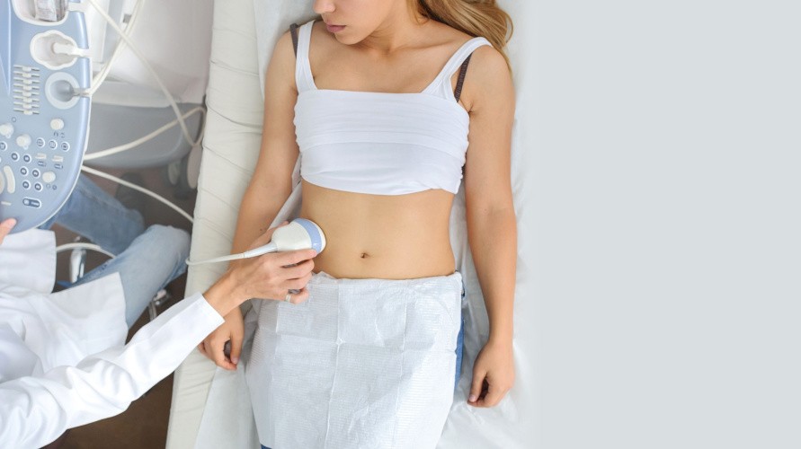
Dokter juga akan menilai kondisi fisik ibu dengan mengukur tanda-tanda vital, seperti tekanan darah dan laju pernapasan untuk mengidentifikasi tanda-tanda syok. Dokter mungkin akan melakukan pemeriksaan fisik dengan melakukan perabaan pada perut atau pemeriksaan ultrasonography (USG) untuk menilai rahim ibu. Jika bagian puncak rahim tidak teraba atau tidak terlihat pada pemeriksaan USG, hal ini dapat menunjukkan inversio uterus.
Pada kasus inversio uterus yang baru terjadi 1 bulan pasca persalinan, penegakan diagnosis sulit dilakukan hanya berdasarkan tanda dan gejala klinis saja. Kasus ini jarang terjadi, dan dapat terjadi hingga 14 minggu setelah persalinan. Gejala yang muncul dapat berupa perdarahan vagina yang persisten, peningkatan suhu tubuh, lemas, dan gejala nyeri pinggang. Untuk diagnosis pasti, perlu dilakukan pemeriksaan USG.
Tata Laksana
Inversio uterus yang terjadi saat persalinan merupakan kondisi gawat darurat dan harus segera diatasi. Pemberian cairan (dan transfusi darah jika diperlukan) harus segera dilakukan untuk menstabilkan fungsi tubuh ibu.
Prosedur Reposisi Manual
Rahim yang mengalami inversi akan dikembalikan ke posisi semula melalui teknik reposisi manual. Teknik itu dilakukan oleh tenaga medis dengan cara mendorong bagian rahim yang keluar menggunakan tangan dan memasukkannya ke dalam. Tindakan ini harus dilakukan segera karena jika ditunda, pengembalian posisi rahim menjadi lebih sulit dan perdarahan yang terjadi akan semakin banyak.
Jika plasenta masih melekat pada rahim, reposisi manual biasanya tidak dilakukan hingga cairan infus diberikan dan obat-obatan yang merelaksasi otot rahim diberikan, seperti nitrogliserin ataupun obat anestesi umum yang dihirup. Dokter juga mungkin akan melakukan prosedur ini di dalam ruang operasi. Antibiotik akan diberikan untuk mencegah terjadinya infeksi. Jika reposisi manual tidak berhasil, operasi dan reduksi hidrostatik dapat dilakukan.
Reposisi Hidrostatik
Reposisi hidrostatik adalah teknik baru yang menggunakan balon yang berisi cairan. Balon akan ditempatkan di rongga rahim dan diisi dengan cairan infus saline untuk mendorong rahim ke posisi normalnya.
Prosedur Operasi
Operasi dapat dilakukan melalui vagina atau dengan membuat sayatan di daerah perut. Pengembalian posisi rahim dilakukan dengan melakukan penarikan jaringan ligamen yang secara normal menjaga posisi rahim di dalam rongga perut. Penarikan ligamen ini dilakukan berulang kali hingga rahim berada pada posisi normal.
Komplikasi
Komplikasi yang terjadi dapat disebabkan karena kondisi inversio uterus itu sendiri (komplikasi primer) atau akibat penatalaksanaannya (komplikasi sekunder).
Komplikasi primer inversio uterus terjadi akibat perdarahan yang ditimbulkannya, di antaranya adalah kerusakan berbagai organ, syok hingga kematian. Jika tidak diobati, perdarahan akan terus terjadi dan jaringan tubuh ibu dapat mengalami nekrosis (kematian jaringan). Kematian juga dapat terjadi jika kondisi ini tidak ditangani segera.
Sementara itu, komplikasi sekunder yang terjadi akibat pengobatan adalah infeksi, kegagalan prosedur dan emboli saline.
Pencegahan
Inversio uterus tidak dapat dicegah, namun dapat ditangani secara efektif bila petugas medis yang menangani Anda bertindak cepat untuk mengatasinya.
Kapan Harus ke Dokter?
Jika Anda melakukan persalinan bukan di rumah sakit, petugas yang menolong persalinan Anda harus segera membawa Anda ke rumah sakit jika tanda-tanda inversio uterus ditemukan.
Mau tahu informasi seputar penyakit lainnya? Cek di sini, ya!
- dr Hanifa Rahma
Cleveland Clinic. (2022). Uterine inversion (inverted uterus): Causes & treatment. Retrieved October 16, 2022, from https://my.clevelandclinic.org/health/diseases/22326-uterine-inversion.
Healthline. (2016, April 07). Pregnancy complications: Uterine inversion. Retrieved October 16, 2022, from https://www.healthline.com/health/pregnancy/complications-uterine-inversion.
Thakur, M., & Thakur, A. (2021). Uterine Inversion. Retrieved October 16, 2022, from https://www.ncbi.nlm.nih.gov/books/NBK525971.
Medscape. (2021, June 26). Malposition of the uterus. Retrieved October 16, 2022, from https://emedicine.medscape.com/article/272497-overview#a6.