Definisi
Tumor retrobulbar atau retro-orbital adalah tumor mata yang terletak di belakang bola mata. Ruang retro-orbital terdiri atas otot mata, saraf, pembuluh darah, dan jaringan lunak. Tumor retro-orbital akan meningkatkan volume di dalam ruang orbita atau rongga mata dan menekan bagian mata lainnya. Tumor retrobulbar juga dapat meluas ke saraf optik, otak, atau sumsum tulang melalui pembuluh darah.
Secara klinis, berkembangnya sel-sel tumor retrobulbar tidak hanya mempengaruhi volume rongga orbita, tetapi juga dapat menimbulkan peradangan, mempengaruhi sistem imun, dan pembesaran sel-sel normal. Proses pembesaran sel-sel tumor di daerah retrobulbar akan menimbulkan penonjolan mata ke luar (proptosis) dan juga ke dalam sehingga menekan saraf optik.
Penyebab
Terdapat beragam jenis tumor retrobulbar, di antaranya:
-
Hemangioma kavernosa
Hemangioma adalah tumor jinak yang dapat tumbuh di dalam rongga orbita. Tumor ini paling sering ditemukan di belakang bola mata dan dapat mendorong mata ke depan sehingga menyebabkan proptosis.
Tumor jinak ini tumbuh lambat dan menimbulkan gejala proptosis yang berkembang perlahan dan tidak disertai nyeri. Sebagian besar tumor bersifat unilateral, atau asimetris pada satu sisi mata. Kasus tumor bilateral (pada kedua mata) lebih jarang terjadi.
-
Glioma
Glioma saraf optik (saraf penglihatan) adalah jenis tumor otak yang tumbuh di sel glia. Sebagian besar glioma saraf optik tidak tumbuh secepat jenis tumor otak lainnya. Glioma banyak ditemukan di persilangan saraf optik kiri dan kanan.
Glioma saraf optik adalah jenis kanker langka yang biasanya tumbuh lambat dan ditemukan pada anak-anak. Kanker ini jarang ditemukan pada usia di atas 20 tahun. Glioma ganas (glioblastoma) lebih jarang terjadi dan hampir selalu pada pria dewasa. Perjalanan penyakit glioblastoma sangat buruk dan hampir pasti meninggal dalam waktu satu tahun. Glioma saraf optik mencapai sekitar 1% dari semua tumor otak.
-
Meningioma
Meningioma adalah tumor yang timbul dari sel arakhnoid pada lapisan meninges, yaitu lapisan pembungkus otak. Tumor ini dapat muncul di rongga orbita sebagai meningioma orbital primer, meningioma yang awalnya berasal dari selubung saraf optik, atau meningioma orbital sekunder, dimana terjadi perluasan dari tumor dari otak ke rongga orbita. Meningioma biasanya tumbuh lambat dan bersifat jinak.
-
Rabdomiosarkoma
Rabdomiosarkoma (RMS) orbital adalah kanker orbital primer yang paling sering terjadi pada anak-anak, yaitu sekitar 35 kasus baru per tahun. Daerah kepala, leher, dan khususnya orbita, merupakan daerah yang sering ditemukan RMS. Tumor ini merupakan tumor ganas yang jika dapat segera didiagnosis, akan menyelamatkan nyawa penderitanya.
RMS orbital terutama merupakan penyakit pada anak-anak, dengan 90% kasus terjadi sebelum usia 16 tahun, dengan rata-rata kasus diderita pada usia 5-7 tahun. Rasio penderita laki-laki dan perempuan adalah 5:3. Tidak ada faktor risiko ras pada kanker ini.
RMS terdiri dari sel-sel otot lurik dalam berbagai tahapan perkembangan yang dapat muncul di beberapa bagian tubuh, termasuk daerah orbita. Kebanyakan RMS orbita muncul di jaringan lunak orbita. Selain itu, RMS juga dapat bersifat sekunder yaitu menyebar dari bagian tubuh lain ke dalam rongga orbita. Kanker ini berkembang di jaringan lunak orbital, bukan di otot ekstraokular.
Faktor Risiko
Hampir sebagian besar tumor bersifat herediter atau diturunkan. Sehingga, risiko Anda lebih tinggi jika memiliki anggota keluarga dengan riwayat tumor mata. Pada hemangioma kavernosa, tidak ditemukan faktor risiko pasti. Namun, kehamilan berkaitan dengan percepatan pertumbuhan tumor yang sudah ada sebelumnya.
Pada meningioma, riwayat terapi radiasi, penyakit neurofibromatosis tipe 2, dan mutasi gen meningkatkan risiko terkena meningioma. Sedangkan pada rabdomiosarkoma, sebagian besar kasus muncul secara spontan. Namun, kanker ini dikaitkan dengan penyakit neurofibromatosis, sindrom Li-Fraumeni, dan malformasi kongenital atau gangguan perkembangan organ seperti pada sindrom Beckwith-Wiedemann.
Gejala
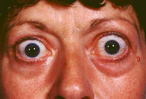
Proptosis atau penonjolan bola mata, yang sering juga disebut dengan eksoftalmus, merupakan gejala khas tumor atau kanker retrobulbar. Proptosis muncul karena adanya pertambahan volume di dalam rongga mata yang mendesak bola mata ke arah luar.
-
Hemangioma kavernosa
Hemangioma kavernosa orbita paling sering terlihat pada wanita paruh baya. Sebagian besar ditemukan di dalam konus otot (rangkaian otot mata berbentuk kerucut), namun bisa juga ditemukan di bagian orbita lainnya. Tumor ini dapat menekan saraf optik sehingga menyebabkan kerusakan.
Terdorongnya bola mata ke depan oleh tumor akan menyebabkan lapisan luar mata kurang terlindungi oleh kelopak mata dan jaringan sekitarnya. Hal ini menyebabkan bola mata menjadi mudah kering, iritasi, bahkan terluka. Penonjolan mata membesar secara perlahan dan tidak nyeri. Gejala penglihatan seperti pandangan ganda, gangguan lapang pandang, gangguan ketajaman penglihatan, dan gangguan fungsi pupil serta otot mata bisa muncul, terjadi akibat penekanan tumor pada jaringan orbital lainnya.
-
Glioma
Gejala glioma saraf optik disebabkan oleh tumor yang menekan saraf. Gejala umum tumor ini meliputi:
-
- mual dan muntah
- masalah keseimbangan
- proptosis bertahap dan tidak nyeri
- gangguan penglihatan
- sakit kepala
- gerakan mata yang tidak disengaja
- gangguan memori
- kantuk siang hari
- kehilangan nafsu makan
- keterlambatan pertumbuhan
- masalah hormon karena tumor terletak dekat bagian otak yang mengatur hormon
- kehilangan penglihatan perlahan
-
Rabdomiosarkoma
Gejala tumor ini umumnya berupa:
-
- proptosis (80-100%) pada satu sisi mata, berkembang pesat dalam beberapa minggu
- pergeseran letak bola mata (80%)
- pembengkakan konjungtiva (selaput bening mata) dan kelopak mata (60%)
- teraba benjolan (25%)
- ptosis atau kelopak mata menutup (25%)
- nyeri dan penurunan penglihatan minimal, merupakan gejala yang lebih lanjut
Diagnosis
-
Hemangioma kavernosa
Hemangioma kavernosa orbita biasanya merupakan tumor yang tumbuh lambat. Jika tumor tidak merusak mata, hemangioma dapat diamati dahulu sebelum mempertimbangkan tindakan intervensi. Pemeriksaan yang dapat dilakukan adalah USG dan CT scan untuk mendeteksi benjolan berbentuk oval atau bulat dan berbatas tegas. Pada pemeriksaan Doppler, akan ditemukan aliran darah yang tenang di dalam tumor.
Pada pemeriksaan jaringan, ditemukan pembesaran pembuluh darah yang terjalin erat. Saluran ini dapat memiliki diameter berukuran 1 mm dan dilapisi oleh satu lapis sel dinding pembuluh darah.
-
Glioma
Pemeriksaan neurologis biasanya akan menunjukkan hilangnya sebagian atau seluruh penglihatan atau perubahan pada saraf optik. Pemeriksaan lain yang digunakan untuk membantu dalam diagnosa glioma saraf optik adalah CT scan, MRI otak, dan biopsi saraf. MRI sering memperlihatkan gambaran pembesaran dan penekukan saraf optik. MRI juga berguna untuk menunjukan penyebaran ke otak.
-
Meningioma
Pada pemeriksaan, dapat ditemukan keluhan kehilangan penglihatan perlahan tanpa nyeri, proptosis, penglihatan ganda, sakit kepala, nyeri belakang mata. Pada pemeriksaan mata, ditemukan saraf optik yang pucat dan bengkak, penurunan tajam penglihatan, gangguan lapang pandang, pergeseran bola mata, dan gerak mata terbatas. Pemeriksaan penunjang yang digunakan adalah pemindairan dengan CT scan dan MRI.
-
Rabdomiosarkoma
Pada pemeriksaan akan ditemukan proptosis, perpindahan bola mata, penonjolan kelopak mata, pembengkakan dan kemerahan kelopak mata, serta teraba benjolan. Tumor pada awalnya tidak mengenai tulang, namun tumor yang lebih besar dapat mengikis tulang dan meluas ke nasofaring (tenggorokan) atau sinus. Pada CT scan, dapat terlihat area perdarahan atau kematian jaringan.
Untuk menunjang diagnosa, dapat dilakukan pemeriksaan biopsi saraf. Biopsi aspirasi jarum halus tidak dapat mencukupi kebutuhan jaringan, sehingga pilihan biopsi biasanya adalah biopsi eksisi atau insisional. Biopsi eksisi dilakukan jika operasi pengangkatan dapat dilakukan tanpa merusak struktur vital mata, seperti saraf dan otot mata. Jika RMS dicurigai besar dan terletak di bagian belakang rongga mata, eksisi lengkap sulit dilakukan tanpa merusak struktur vital orbital, biopsi insisional bisa menjadi pilihan.
Tata Laksana
Pada kanker mata retrobulbar, dapat dilakukan prosedur pembedahan dengan berbagai macam metode, di antaranya:
- Orbitotomi, yaitu pengangkatan tumor dengan membuka jaringan orbita
- Enukleasi, yaitu pengangkatan seluruh bola mata
- Eksenterasi orbita, yaitu pengambilan seluruh atau sebagian jaringan
- Pembedahan endoskopi endonasal, yaitu operasi menggunakan kamera kecil melalui lubang hidung
Pilihan terapi lainnya adalah kemoterapi. Pada kemoterapi kanker mata retrobulbar, biasanya pemberian obat dilakukan melalui suntikan ke pembuluh darah, diberikan selama tiga sampai enam siklus.
Komplikasi
Beberapa komplikasi tumor retrobulbar maupun efek terapinya antara lain:
- Kehilangan penglihatan
- Cedera saraf dan otot mata pasca operasi
- Penyebaran kanker ke bagian tubuh lain, membuat pengobatan dan pemulihan semakin lama. Penyebaran tersering dari rabdomiosarkoma adalah ke paru, kelenjar getah bening, dan tulang
- Efek samping kemoterapi seperti mual, muntah, rambut rontok, lemas, dan lain-lain
Pencegahan
Belum ada cara pasti untuk mencegah tumor atau kanker mata, termasuk tumor retrobulbar. Namun, terdapat bukti adanya hubungan antara sinar matahari dengan kanker. Sehingga, hal yang dapat dilakukan untuk mengurangi risikonya adalah dengan membatasi paparan matahari ke mata menggunakan topi atau kacamata hitam dengan pelindung sinar ultraviolet. Selan itu, membatasi paparan zat kimia ke tubuh, terutama pada saat hamil juga dapat mencegah munculnya mutasi gen akibat bahan kimia.
Kapan Harus ke Dokter?
Segera periksa ke dokter jika Anda mengalami tanda dan gejala tumor retrobulbar seperti yang disebutkan di atas. Terutama jika terdapat proptosis dan tidak ditemukan penyebab lainnya.
Mau tahu informasi seputar penyakit lainnya? Cek di sini, ya!
- dr Hanifa Rahma
Cohen, A. (2021). Orbital cavernous hemangioma. Retrieved 26 Desember 2021, from https://emedicine.medscape.com/article/1218120-overview
Herndon, J. (2016). Optic nerve glioma. Retrieved 26 Desember 2021, from https://www.healthline.com/health/optic-glioma
Fowler, B., Burkat, C., and Wilson, M. (2021). Rhabdomyosarcoma. Retrieved 26 Desember 2021, from https://eyewiki.aao.org/Rhabdomyosarcoma
Fowler, B., Kim, J. (2021). Orbital meningiomas. Retrieved 26 Desember 2021, from https://eyewiki.org/Orbital_Meningiomas
Soebagjo, H. (2019). Onkologi mata. Pusat Penerbitan dan Percetakan Universitas Airlangga: Surabaya
Finger, P. (2021). Cavernous hemangioma of the orbit. Retrieved 26 Desember 2021, from https://eyecancer.com/eye-cancer/conditions/orbital-tumors/cavernous-hemangioma-of-the-orbit/
Pargament, J., Nerad, J. (2021). Orbital Cavernous Venous Malformation (Cavernous Hemangioma). Retrieved 26 Desember 2021, from https://eyewiki.aao.org/Orbital_Cavernous_Venous_Malformation_(Cavernous_Hemangioma)#Complications
Alsuhaibani, A. (2021). Optic nerve glioma. Retrieved 26 Desember 2021, from https://eyewiki.aao.org/Optic_Nerve_Glioma
American Cancer Society (2018). Can eye cancer be prevented?. Retrieved 26 Desember 2021, from https://www.cancer.org/cancer/eye-cancer/causes-risks-prevention/prevention.html